live_tv
Livestream Starting Soon
00
Hours
:
00
Minutes
:
00
Seconds
Up next in 10

Facilitated Diffusion - Unlocking Cell Transport
In this educational video, we delve into the fascinating process of facilitated diffusion, a crucial mechanism in cellular transport. Discover how molecules move across cell membranes with the help of specific transport proteins, ensuring that essential nutrients and ions enter and exit cells efficiently. We will explore the differences between facilitated diffusion and other transport methods, such as passive and active transport, and highlight the significance of this process in maintaining cellular homeostasis. Join us as we unlock the secrets of cell transport and its vital role in biological systems. #FacilitatedDiffusion #CellTransport #Biology
Website: https://biologynotesonline.com/
Facebook: https://www.facebook.com/biologynotesonline
Instagram: https://www.instagram.com/biologynotesonline/?hl=en
facilitated diffusion,diffusion,membrane transport,active transport,transport,transport in cells diffusion and osmosis,facilitated diffusion a level biology,aqa a level biology facilitated diffusion,facilitated membrane transport,transport across cell membranes: diffusion,cell membrane transport,passive transport,transport in cells,active transport in cells,passive transport in cells,cell transport,transport across cell membrane
Show More Show Less View Video Transcript
0:00
facilitated diffusion is a critical transport mechanism that allows certain molecules to move across cell
0:07
membranes Cell membranes form a protective barrier around cells These membranes contain specialized transport
0:14
proteins that create pathways through the phospholipid billayer Facilitated diffusion always moves substances from
0:22
areas of higher concentration to areas of lower concentration Unlike active transport
0:29
facilitated diffusion doesn't require energy The molecules naturally move down their concentration
0:43
gradient Facilitated diffusion is essential for transporting molecules that cannot pass through the
0:49
phospholipid blayer on their own such as water- soluble molecules ions glucose
0:54
and amino acids The cell membrane forms a critical
1:00
barrier that separates the internal environment of the cell from the external
1:05
surroundings This membrane is composed of a phosphoipid blayer with two layers of phospholipids arranged in opposite
1:12
directions Each phospholipid has a hydrophilic head that interacts with water and hydrophobic tails that avoid
1:19
water The membrane acts as a selective barrier allowing some molecules to pass through
1:25
while blocking others Small non-polar molecules like oxygen and carbon dioxide
1:31
can easily diffuse through the hydrophobic core of the membrane Small polar molecules like water can pass
1:37
through but at a slower rate due to the hydrophobic nature of the membrane interior Large molecules however cannot
1:44
pass through the membrane due to their size Charged molecules like ions are
1:49
repelled by the hydrophobic environment and cannot easily cross without assistance Despite the membrane barrier
1:57
cells need to transport essential molecules like glucose amino acids and ions This selective permeability creates
2:04
the need for facilitated diffusion mechanisms specialized transport proteins that help essential molecules
2:11
cross the membrane barrier
2:25
Concentration gradients are fundamental to understanding facilitated diffusion A
2:30
concentration gradient exists when the concentration of molecules differs between two adjacent areas
2:38
Let's visualize a cell membrane with a high concentration of molecules on one side and a low concentration on the
2:44
other The membrane contains transport proteins that allow specific molecules to pass through in a process called
2:50
facilitated diffusion According to the concentration gradient molecules naturally move from areas of high
2:57
concentration to areas of low concentration This movement follows the second law of thermodynamics which
3:04
states that systems tend toward maximum entropy or disorder A key feature of
3:09
facilitated diffusion is that it requires no energy input The movement is driven by the concentration gradient
3:16
alone making it a passive transport process Transport proteins are the key
3:23
components that enable facilitated diffusion across cell membranes The cell membrane consists of a phospholipid
3:30
billayer that forms a barrier to most molecules Transport proteins have several important features that allow
3:37
them to facilitate the movement of specific molecules These specialized proteins are
3:43
embedded directly within the cell membrane Each transport protein has specific binding sites that recognize
3:50
and bind to particular molecules After binding transport proteins undergo a
3:55
confirmational change altering their shape to move substances across the membrane This elegant mechanism allows
4:02
molecules to pass through the membrane without disrupting its structural integrity which is crucial for cell
4:09
survival Transport proteins are essential components that enable cells to precisely control which molecules can
4:16
enter and exit making facilitated diffusion possible Channel proteins are specialized
4:22
transport proteins that create water-filled passageways through the cell membrane Unlike carrier proteins channel
4:30
proteins form permanent pores or tunnels that allow molecules to flow through the membrane Channel proteins are highly
4:37
selective allowing only specific types of molecules to pass through This selectivity is based primarily on the
4:44
size of the molecules Small molecules can pass through while larger ones cannot Additionally many channels are
4:51
selective based on the electrical charge of molecules Let's look at two important examples of channel proteins found in
4:58
cell membranes Aquaporins are specialized channel proteins that allow water molecules to pass through the
5:04
membrane efficiently These channels are so efficient that billions of water molecules can pass through a single
5:11
aquaporn each second Ion channels are another important type
5:16
of channel protein that allow specific ions to pass through the membrane These
5:22
channels are highly selective for specific ions such as potassium sodium calcium or chloride For example a
5:28
potassium channel allows potassium ions to pass while blocking other ions It's
5:33
important to note that many channel proteins are not always open They can be regulated by gates that respond to
5:40
different stimuli Carrier proteins facilitate the movement of specific molecules across the cell
5:47
membrane through a unique mechanism Unlike channel proteins carrier proteins
5:52
undergo significant confirmational changes during transport Let's examine how glucose transporters or glut
5:59
proteins move glucose across the membrane First the carrier protein has a specific binding site that recognizes
6:06
glucose molecules After glucose binds the carrier protein underos a
6:11
confirmational change rotating to expose the binding site to the opposite side of the membrane The glucose molecule is
6:18
then released into the intracellular environment due to the lower affinity of the binding site in this new
6:25
confirmation Finally the empty carrier protein returns to its original confirmation ready to transport another
6:32
glucose molecule Glut proteins are a family of glucose transporters with several key
6:38
features Let's compare simple diffusion and facilitated diffusion two fundamental processes for moving
6:45
molecules across cell membranes Simple diffusion occurs when molecules move directly through the phospholipid
6:51
billayer without any assistance from proteins In contrast facilitated
6:56
diffusion requires specialized transport proteins embedded in the membrane to help molecules cross Simple diffusion is
7:03
limited to small non-polar molecules like oxygen and carbon dioxide that can
7:08
slip between the lipid tails of the membrane Facilitated diffusion however can transport larger or polar molecules
7:16
like glucose and amino acids that cannot pass directly through the hydrophobic
7:21
core of the membrane Simple diffusion doesn't require proteins and is limited to small non-polar molecules Its rate is
7:28
directly proportional to the concentration gradient Facilitated diffusion requires transport proteins
7:34
and accommodates larger or polar molecules It works faster than simple diffusion but can become saturated when
7:42
all transport proteins are in use When we compare their transport rates simple diffusion shows a linear relationship
7:49
with concentration While facilitated diffusion demonstrates a saturation curve as transport proteins become fully
7:56
occupied To summarize both types of diffusion move molecules from high to low concentration without energy input
8:03
but they differ in speed specificity and the types of molecules they
8:09
transport Let's compare facilitated diffusion and active transport two fundamental mechanisms for moving
8:16
molecules across cell membranes Facilitated diffusion is a passive transport process that moves molecules
8:22
from areas of higher concentration to lower concentration through specialized transport
8:27
proteins In facilitated diffusion molecules move down their concentration gradient without requiring energy The
8:35
key characteristics of facilitated diffusion are it's a passive process requiring no energy It follows the
8:41
concentration gradient and it uses transport proteins to help molecules cross the membrane
8:47
In contrast active transport is a process that moves molecules against their concentration gradient using
8:53
energy typically in the form of ATP Active transport pumps use energy from
8:58
ATP to move molecules against their concentration gradient from areas of lower concentration to areas of higher
9:05
concentration The key characteristics of active transport are it's an active process requiring energy from ATP It
9:13
works against the concentration gradient and it uses specialized transport
9:19
pumps Let's compare these two transport mechanisms side by side to better understand their key differences While
9:26
facilitated diffusion requires no energy active transport depends on ATP as an energy source Facilitated diffusion
9:34
always moves molecules down their concentration gradient Whereas active transport can move molecules against
9:40
their gradient The rate of facilitated diffusion is limited by the strength of the concentration gradient while active
9:47
transport can achieve higher transport rates Both processes show saturation
9:52
kinetics meaning they can reach a maximum rate when all transport proteins are occupied Facilitated diffusion is
9:59
commonly used for glucose and amino acid transport while active transport includes examples like the sodium
10:06
potassium pump and calcium pumps The fundamental difference between these two transport mechanisms lies in their
10:13
energy requirements and their relationship to concentration gradients Facilitated diffusion relies
10:20
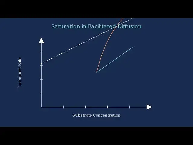
on transport proteins to move molecules across cell membranes Saturation is a
10:26
key property that differentiates facilitated diffusion from simple diffusion It occurs when all available
10:32
transport proteins are actively engaged in moving substrates At low substrate
10:38
concentration only a few transport proteins are engaged in moving molecules As substrate concentration increases
10:45
more transport proteins become occupied At high substrate concentration all transport proteins are engaged leading
10:52
to maximum transport rate Let's visualize this with a graph showing transport rate versus substrate
11:00
concentration In facilitated diffusion the transport rate initially increases with substrate concentration But as
11:07
substrate concentration continues to increase we reach a point of saturation where all transport proteins are
11:14
occupied This creates a maximum transport rate that cannot be exceeded even if we further increase substrate
11:20
concentration In contrast simple diffusion doesn't rely on transport proteins and shows a linear relationship
11:27
without saturation To summarize saturation is a defining characteristic of facilitated
11:34
diffusion It occurs when all transport proteins are engaged creating a maximum
11:39
transport rate that cannot be exceeded This distinguishes it from simple diffusion which shows a linear
11:46
relationship between concentration and transport rate without saturation
11:51
Glucose transport across cell membranes is a classic example of facilitated diffusion in action Glucose is a vital
11:58
energy source for cells but it faces a challenge It's a relatively large polar
12:03
molecule that cannot cross the hydrophobic cell membrane on its own Without assistance glucose would be
12:09
unable to enter cells efficiently despite the concentration gradient driving it
12:15
inward This is where glut proteins come in Glitz stands for glucose transporter
12:21
a family of specialized transmembrane proteins that create channels specifically for glucose to pass
12:27
through The transport mechanism involves a series of steps First glucose binds to
12:33
a specific site on the glut protein This causes the protein to change shape moving the glucose through the membrane
12:40
Finally glucose is released into the cell and the protein returns to its original confirmation
12:47
Different cell types have specialized glit proteins based on their glucose needs Glute one is abundant in red blood
12:54
cells and the brain ensuring a constant supply of glucose Glute 2 in the liver
12:59
and pancreas helps with glucose sensing Glute 4 found in muscle and fat cells is
13:05
unique because it responds to insulin moving to the cell membrane only when insulin is present
13:12
The efficient transport of glucose is critical for cellular energy production Once inside the cell glucose enters the
13:19
glycolysis pathway eventually producing ATP the energy currency of cells This
13:25
energy powers everything from muscle contraction to neuron firing to cell
13:30
division Understanding glucose transport has important clinical implications In
13:35
diabetes glut 4 proteins fail to respond properly to insulin preventing glucose
13:41
uptake in muscle and fat tissues Glut one deficiency can cause seizures and developmental delays due to insufficient
13:48
glucose in the brain Cancer cells often increase glut expression to fuel their rapid growth
13:55
In summary glucose transport via glip proteins is a perfect illustration of facilitated diffusion in action
14:01
demonstrating how cells have evolved elegant solutions to move essential molecules across membrane barriers Ion
14:08
channels are specialized membrane proteins that facilitate the diffusion of specific ions across the cell
14:14
membrane These channels are highly selective allowing only specific ions such as sodium potassium or calcium to
14:22
pass through Many ion channels are gated meaning they can open and close in
14:27
response to specific stimuli There are three main types of gated channels Voltage gated channels respond to
14:34
electrical signals Lean gated channels respond to chemical messengers And mechanically gated channels respond to
14:41
physical forces One of the most important examples of
14:46
ion channels in action is the generation of action potentials in neurons Neurons
14:51
contain voltage gated sodium and potassium channels that open and close at different times to generate
14:57
electrical signals During an action potential sodium channels open first
15:03
allowing sodium ions to rush into the cell causing depolarization Then potassium channels open allowing
15:09
potassium ions to exit the cell causing repolarization and a return to the resting state To summarize ion channels
15:16
are specialized proteins that facilitate the diffusion of specific ions across cell membranes They're critical for
15:23
neuronal signaling muscle contraction and many other biological
15:29
functions Several key factors influence the rate of facilitated diffusion across cell membranes Let's examine the four
15:37
main factors that determine how quickly molecules move through transport proteins The steepness of the
15:44
concentration gradient is a primary factor affecting diffusion rate A steeper concentration gradient meaning a
15:51
greater difference in molecule concentration between the two sides of the membrane results in faster
15:58
diffusion The second factor is the number of transport proteins in the membrane Membranes with more transport
16:04
proteins allow for faster facilitated diffusion With more proteins more molecules can pass through
16:11
simultaneously increasing the overall rate of diffusion Temperature is the third major
16:17
factor affecting facilitated diffusion rates Higher temperatures increase the
16:22
kinetic energy of molecules causing them to move more rapidly This increased
16:27
molecular motion leads to more frequent collisions with transport proteins accelerating the rate of facilitated
16:35
diffusion The fourth factor is molecular specificity which refers to how well molecules match the specific transport
16:43
proteins Transport proteins are highly selective only allowing molecules with
16:48
the correct size and shape to pass through Molecules that match the transport proteins binding site can pass
16:54
through while others cannot regardless of the concentration gradient To summarize four main factors
17:02
determine the rate of facilitated diffusion across cell membranes First the steepness of the concentration
17:08
gradient drives the direction and speed of diffusion Second the number of transport proteins in the membrane
17:15
determines how many molecules can pass through simultaneously Third temperature affects molecular
17:21
kinetic energy and movement speed And fourth molecular specificity ensures only compatible molecules can utilize
17:28
specific transport proteins Understanding these factors helps explain how cells regulate the movement
17:35
of essential molecules across their membranes Temperature significantly affects how quickly molecules move
17:42
through transport proteins in facilitated diffusion At normal body temperature around 37 degrees C
17:49
facilitated diffusion occurs at an optimal
18:01
rate When temperature decreases molecular movement slows down Lower temperatures reduce the kinetic energy
18:08
of molecules and decrease protein flexibility
18:22
As temperature increases molecules move faster and transport proteins become more flexible This increases the rate of
18:29
facilitated diffusion
18:38
However at extreme temperatures above 50° C proteins begin to denature Protein
18:44
dennaturation alters the three-dimensional structure of transport proteins causing the channel to collapse
18:50
and preventing molecules from passing through
19:00
This graph shows how temperature affects the rate of facilitated diffusion The rate peaks at normal body temperature
19:07
around 37° At low temperatures the transport rate is reduced due to decreased
19:13
molecular movement and protein flexibility At optimal temperature facilitated diffusion reaches its
19:19
maximum rate As temperature increases above the optimum the rate initially
19:24
increases but then begins to decline as proteins start to destabilize At extreme
19:29
temperatures protein dennaturation occurs causing facilitated diffusion to essentially stop To summarize
19:36
temperature affects facilitated diffusion in multiple ways Higher temperatures increase molecular movement
19:42
and make transport proteins more flexible both of which enhance diffusion rates
19:47
However extreme temperatures denature proteins and stop the transport process
19:53
completely PH plays a crucial role in facilitated diffusion by affecting the structure and function of transport
20:00
proteins Most transport proteins have an optimal pH range where they function most efficiently This is typically
20:07
around neutral pH for many cellular proteins At optimal pH the protein
20:12
structure allows molecules to pass through efficiently via facilitated diffusion Transport efficiency follows a
20:19
bell curve relationship with pH with maximum efficiency at the protein's optimal pH range When pH changes it
20:27
affects the ionization state of amino acids in the protein altering hydrogen bonds and electrostatic interactions In
20:34
acidic environments the increased concentration of hydrogen ions can protonate negatively charged amino acids
20:40
changing the protein shape PH changes directly affect binding sites by altering their shape and
20:47
chemical properties Acidic or basic conditions can disrupt the precise configuration needed for substrate
20:54
recognition In basic environments hydroxide ions can deproinate positively
21:00
charged groups again disrupting the protein structure and function Outside of the optimal pH range transport
21:07
efficiency decreases significantly This is why maintaining proper pH in cellular
21:12
compartments is essential for normal function Understanding pH effects on transport proteins is critical in both
21:18
normal physiology and in pathological conditions where pH balance is disrupted
21:25
Transport proteins are essential for proper cellular function allowing specific molecules to cross the cell
21:32
membrane through facilitated
21:37
diffusion When transport proteins malfunction serious medical conditions can result We'll examine three
21:43
significant disorders Glucose galactose malabsorption cystic fibrosis and
21:49
certain forms of diabetes Glucose galactose malabsorption is caused by mutations in the SGLT1
21:55
transporter In healthy intestines SGLT1 facilitates absorption of glucose and
22:01
galactose from food With defective SGLT1 transporters these sugars cannot be
22:06
absorbed The resulting buildup of sugars in the intestine causes severe diarrhea and dehydration particularly in infants
22:15
Cystic fibrosis results from mutations in the CFTR gene which normally creates
22:20
chloride ion channels in cell membranes These channels are critical for maintaining proper fluid balance When
22:27
CFTR channels malfunction chloride ions cannot pass through properly This leads
22:33
to the production of thick sticky mucus that clogs airways in the lungs and obstructs the pancreas and other organs
22:40
In diabetes mellus particularly type 2 diabetes cells develop insulin resistance Normally insulin triggers the
22:48
movement of glut glucose transporters to the cell membrane With insulin
22:53
resistance this glute 4 transllocation is impaired preventing efficient glucose uptake from the bloodstream This leads
23:00
to chronically elevated blood sugar levels and resulting metabolic complications
23:06
Understanding the molecular basis of transport protein disorders has led to targeted treatment approaches For
23:12
glucose galactose malabsorption dietary management is essential eliminating glucose and galactose from the diet For
23:19
cystic fibrosis breakthrough CFTR modulator drugs have been developed that can improve protein folding trafficking
23:26
and function for specific mutations addressing the root cause of the disease
23:32
In diabetes medications like thoazoladine do act as insulin sensitizers improving glute 4
23:38
transllocation to the cell membrane and enhancing glucose
23:43
uptake Scientists use specialized laboratory techniques to study facilitated diffusion across cell
23:51
membranes The first major technique is radioactive tracers In this method
23:56
molecules are labeled with radioactive isotopes such as tridium or carbon 14
24:02
This allows scientists to track their movement across membranes and precisely measure transport
24:07
rates The second technique is fluorescent tagging Transport proteins are labeled with fluorescent molecules
24:14
like green fluorescent protein Using fluorescent microscopy scientists can
24:19
visualize protein locations and track their movements in real time
24:24
The third key technique is patch clamping A glass micro pipet forms a tight seal with a small patch of cell
24:32
membrane This allows scientists to record electrical currents as ions move through individual channel proteins This
24:39
technique provides detailed functional data about how transport channels operate Scientists also use advanced
24:46
imaging techniques Methods like fret can measure interactions between proteins Total internal reflection fluoresence or
24:54
turf visualizes molecules near the membrane surface Cryeleron microscopy
24:59
reveals detailed threedimensional structures of transport proteins These experimental methods have
25:06
profoundly advanced our understanding of facilitated diffusion They've revealed detailed structures of transport
25:13
proteins identified key binding sites and established connections between
25:18
protein structure and function This knowledge has also been crucial for developing treatments for diseases
25:25
related to transport disorders Plant cells employ facilitated
25:31
diffusion for essential molecular transport across their membranes Plant cells utilize specialized channel
25:37
proteins called aquaporins that facilitate water movement across their membranes
25:45
Aquaporins increase membrane permeability to water allowing efficient hydration and maintaining tur pressure
25:52
which is crucial for plant structural support Plant cells also employ carrier
25:58
proteins for nutrient uptake such as glucose amino acids and essential
26:03
minerals These carrier proteins facilitate the selective transport of nutrients down their concentration
26:09
gradients without requiring energy Let's compare facilitated diffusion in
26:16
plant versus animal cells Plant cells have unique adaptations for facilitated diffusion They require transport across
26:24
cell walls use vacules for storage and employ plasma for cellto cell transport
26:30
To summarize plant cells rely on facilitated diffusion for water and nutrient transport through specialized
26:37
proteins Despite structural differences from animal cells they share similar transport protein mechanisms while
26:44
addressing plant specific requirements
27:35
Cellular respiration is a vital process that converts glucose into energy But for this to occur molecules must first
27:42
enter the cell and move between different cellular compartments Facilitated diffusion plays
27:48
a crucial role right from the start of cellular respiration Glucose the primary energy source must first enter the cell
27:56
Glucose molecules cannot easily pass through the cell membrane's phospholipid billayer Instead they rely on specific
28:03
G-let transporters embedded in the membrane Once glucose enters the cell it
28:09
underos glycolysis in the cytoplasm to form pyuvate For cellular respiration to
28:14
continue pyrovate must enter the mitochondria Pyrovate molecules use specific mitochondrial pyrovate carriers
28:21
or MPCs to cross the outer mitochondrial membrane through facilitated
28:26
diffusion Beyond glucose and pyrovate cellular respiration involves the transport of many other molecules
28:33
through facilitated diffusion ADP and ATP move in and out of mitochondria via
28:39
the adinine nucleotide translocator Fatty acids another energy source enter mitochondria through the carnitine
28:45
shuttle system and electrons move through protein complexes in the electron transport chain These transport
28:52
systems must be coordinated to maintain efficient cellular respiration The rate of glucose entry must match metabolic
28:59
demands Glucose enters the cell pyuvate moves into mitochondria and ATP is
29:05
transported out to power cellular activities This coordination ensures that energy production meets cellular
29:11
needs Transport rates adjust to metabolic demands and regulatory mechanisms control protein activity
29:18
Defects in any of these transport systems can disrupt energy production and lead to metabolic
29:25
disorders Computational modeling has revolutionized our understanding of facilitated diffusion by allowing
29:31
scientists to visualize and analyze transport processes at the atomic level
29:36
Scientists use several computational approaches to model facilitated diffusion Molecular dynamics simulates
29:43
atomic movements Monte Carlo methods model probabilistic events and machine
29:48
learning predicts protein function from structural data These computational models reveal critical insights into the
29:55
relationship between a transport protein structure and its function They help identify binding sites channel
30:02
mechanisms and confirmational changes that facilitate diffusion For example a model can show
30:08
how a molecule binds to a specific site passes through the channel and underos confirmational changes during
30:15
transport Let's examine a computational simulation of facilitated diffusion
30:20
across a membrane The model tracks molecules moving from an area of high concentration to low concentration
30:27
through a specific channel protein The simulation calculates how molecules interact with the channel protein based
30:33
on physical properties like molecular size charge and the specific shape of the channel These computational models
30:40
have wide ranging applications in research and development from drug design to disease modeling and
30:46
biotechnology Despite advances challenges remain in improving computational accuracy
30:52
integrating models across multiple scales and validating predictions with experimental data As computational power
31:00
increases these models will continue to provide deeper insights into the mechanisms of facilitated diffusion
31:07
Research on facilitated diffusion continues to advance in several exciting directions
32:09
One major research frontier is the determination of transport protein structures Advanced techniques like
32:16
cryeleron microscopy are revealing how these proteins change shape during transport
32:24
Personalized medicine is another exciting frontier Researchers are studying how genetic variations in
32:30
transport proteins affect drug response enabling tailored treatment
32:36
approaches The development of artificial transport systems is a growing field
32:41
Scientists are creating synthetic membranes with engineered channels that mimic biological transport proteins
32:51
These research directions will have significant impacts on medicine and biotechnology from improved drug
32:57
delivery systems to novel diagnostic technologies and treatment approaches As these interconnected research areas
33:04
advance we're gaining deeper insights into facilitated diffusion and developing innovative applications that
33:10
could transform medicine and biotechnology Facilitated diffusion is a critical
33:16
process that allows specific molecules to cross cell membranes without using cellular energy This passive transport
33:23
process enables molecules to move down their concentration gradient through specialized transport
33:30
proteins Facilitated diffusion relies on two main types of transport proteins channel proteins and carrier proteins
33:38
Channel proteins form water- fil pores that allow specific ions or molecules to pass through and can be always open or
33:45
gated Carrier proteins bind specific molecules and undergo confirmational
33:51
changes to transport them across the membrane Facilitated diffusion has
33:56
several key characteristics that distinguish it from other transport mechanisms It's a passive process
34:02
requiring no energy Follows concentration gradients demonstrates high specificity for molecules exhibits
34:08
saturation kinetics and is sensitive to environmental factors like temperature and
34:13
pH The biological significance of facilitated diffusion cannot be overstated It's essential for nutrient
34:20
uptake ion homeostasis cell signaling water balance and even has implications
34:26
for drug delivery and therapeutic design In conclusion facilitated diffusion is
34:31
fundamental to life enabling cells to maintain their internal environment while exchanging materials with their
34:39
surroundings Facilitated diffusion serves as the essential bridge between cells and their environment allowing
34:46
life as we know it to exist
#Health
#Biological Sciences