0:00
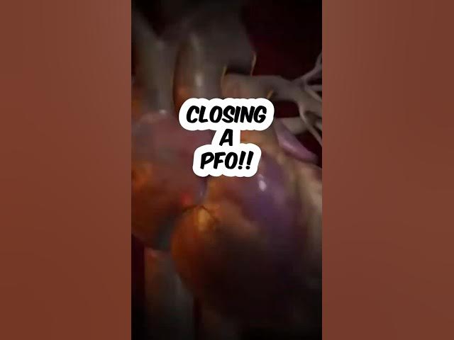
So this is an animation of the heart and
0:03
you can see now as they cut into the
0:05
heart this wire is coming up from the
0:07
right side and opening the flap of the
0:09
PFO and it shows nicely the nature of a
0:12
PFO being this flap that can be pushed
0:14
open and then the device is now opened
0:17
in the left brought down to the septum
0:20
and another identical disc is brought up
0:24
on the right side. The two are locked
0:26
together by this nitonol memory metal
0:28
loop and then that clamps close the PFO
0:32
and now they're animating some pink
0:35
cells growing over the device and that's
0:38
the process of what we call
0:39
endothelialization or healing up of the
0:42
septum and the cells are using the
0:45
device as a scaffold over which to grow
0:48
across which is what should have
0:49
happened in uh when when the person was
0:53
a baby to close off the PFO.