Up next in 10


Molecular Diagnosis of Genetic Diseases
Website: https://biologynotesonline.com/
Facebook: https://www.facebook.com/biologynotesonline
Instagram: https://www.instagram.com/biologynotesonline/?hl=en
Show More Show Less View Video Transcript
0:00
Welcome to our introduction to the
0:01
molecular diagnosis of genetic diseases.
0:04
In this video, we'll explore how
0:06
molecular techniques have revolutionized
0:08
the identification and management of
0:11
genetic disorders.
0:14
Molecular diagnosis is the process of
0:16
identifying diseases by studying
0:17
molecules such as DNA, RNA, and
0:20
proteins. This approach allows us to
0:22
detect genetic abnormalities at the
0:24
molecular level, providing precise
0:26
information about the genetic basis of
0:29
diseases.
0:31
Molecular diagnostics has revolutionized
0:34
medicine over the past few decades,
0:36
starting in the 1970s with early DNA
0:39
sequencing methods. The 1980s saw the
0:42
first diagnostic tests for genetic
0:44
disorders, followed by the human genome
0:46
project in the 1990s, the complete
0:49
sequencing of the human genome in the
0:52
2000s to today's rapid and affordable
0:55
genetic testing technologies. Before
0:57
molecular diagnostics, identification of
0:59
genetic diseases relied primarily on
1:02
symptoms and family history. Now we can
1:04
precisely identify specific genetic
1:06
mutations and varants enabling earlier
1:09
and more accurate diagnosis.
1:12
Molecular diagnosis offers several key
1:15
benefits in the management of genetic
1:16
diseases. It enables early detection of
1:19
disease predisposition often before
1:21
symptoms appear. It allows precise
1:24
classification of disease subtypes which
1:26
can vary in severity and progression.
1:29
This precision helps in selecting
1:31
personalized treatments based on
1:33
specific genetic varants. It facilitates
1:36
family screening and genetic counseling
1:38
for relatives who may be at risk. And it
1:41
provides a way to monitor disease
1:42
progression and response to treatment
1:45
over time. Early and accurate diagnosis
1:47
is critical for optimal disease
1:49
management and improved patient
1:51
outcomes.
1:53
In this video series, we'll focus on
1:56
three major genetic diseases as case
1:58
studies to understand molecular
2:00
diagnostic approaches. First, we'll
2:02
explore cystic fibrosis, a condition
2:05
affecting the respiratory and digestive
2:07
systems. Next, Huntington's disease, a
2:10
progressive neurodeenerative disorder.
2:12
And finally, cickle cell anemia, which
2:14
affects red blood cells and oxygen
2:16
transport throughout the body. For each
2:18
disease, we'll examine how molecular
2:20
diagnostic techniques can identify
2:22
specific genetic mutations. In cystic
2:25
fibrosis, the most common mutation is a
2:28
deletion of three nucleotides.
2:30
Huntington's disease is characterized by
2:32
an expansion of KAG repeats, and cickle
2:35
cell anemia results from a single
2:37
nucleotide substitution. Understanding
2:39
these specific mutations is key to
2:41
accurate diagnosis and developing
2:44
targeted treatments for genetic
2:45
diseases.
2:48
Molecular diagnostics is based on
2:50
detecting specific DNA or RNA sequences
2:53
associated with genetic diseases. There
2:55
are four key components to molecular
2:57
diagnostic techniques. First, these
3:00
methods target DNA or RNA molecules, the
3:03
genetic material that carries the
3:04
blueprint for all cellular functions.
3:07
Second, they rely on base pair
3:08
complimentarity where adinine pairs with
3:10
thymine and guanine pairs with cytosine.
3:14
Third, they offer sequence specificity,
3:16
allowing detection of exact genetic
3:18
sequences linked to diseases. Finally,
3:21
most techniques include amplification
3:23
steps to make even small amounts of
3:25
genetic material detectable.
3:28
Molecular diagnostic methods detect
3:30
specific DNA or RNA sequences through a
3:33
process of targeted binding. Probes with
3:36
complimentary sequences bind only to
3:38
their exact matching targets through
3:40
base pairing. These probes only bind to
3:43
their exact complimentary sequences,
3:45
providing the specificity needed to
3:47
identify disease-causing mutations. When
3:50
binding occurs, various detection
3:52
methods convert this molecular
3:54
recognition event into measurable
3:55
signals that can be analyzed.
3:59
Let's compare molecular diagnostics with
4:01
traditional diagnostic approaches.
4:03
Traditional diagnostics focuses on
4:06
symptoms and provides indirect evidence
4:08
of disease, while molecular diagnostics
4:10
targets the actual genetic cause through
4:12
direct DNA or RNA detection. Molecular
4:16
methods often enable earlier detection
4:18
before symptoms appear and provide more
4:21
precise genetic identification compared
4:23
to the broader diagnostic categories of
4:25
traditional approaches. Perhaps most
4:28
importantly, molecular diagnostics
4:30
guides more personalized treatment
4:32
options based on a patient's specific
4:34
genetic profile. The key difference is
4:37
that molecular diagnostics identifies
4:39
the exact genetic cause, not just the
4:42
clinical manifestations of disease. By
4:44
targeting the molecular basis of
4:46
disease, these techniques provide
4:48
precision that transforms our approach
4:51
to diagnosis, treatment, and prevention
4:53
of genetic disorders.
4:56
Molecular diagnostic techniques allow us
4:58
to analyze DNA and genetic variations at
5:01
the molecular level. The four primary
5:04
techniques are PCR, DNA sequencing, fish
5:06
and microarray analysis.
5:09
Polymerase chain reaction or PCR is a
5:11
technique used to amplify specific DNA
5:13
segments. The PCR process involves three
5:16
main steps. Dennaturation, analing, and
5:18
extension.
5:20
PCR has numerous applications including
5:23
pathogen detection, identification of
5:25
genetic mutations, and gene expression
5:27
analysis.
5:29
DNA sequencing determines the precise
5:31
order of nucleotides in a DNA molecule.
5:34
This process can be performed using
5:36
traditional Sanger sequencing or newer
5:38
next generation sequencing methods.
5:41
DNA sequencing is essential for
5:43
identifying genetic varants, diagnosing
5:46
genetic disorders, and studying
5:47
evolutionary relationships.
5:50
Fluorescent and C2 hybridization or fish
5:53
uses fluorescent probes to detect
5:55
specific DNA sequences on chromosomes.
5:58
In this technique, fluorescently labeled
5:59
DNA probes bind to complimentary
6:01
sequences on chromosomes, allowing
6:03
visualization of specific genetic
6:05
regions. The fish process involves
6:08
denaturing DNA, hybridizing fluorescent
6:11
probes, washing away unbound probes, and
6:13
visualizing under a fluorescent
6:15
microscope. Fish is valuable for
6:17
detecting chromosomeal abnormalities,
6:20
identifying gene amplifications or
6:21
deletions, and locating specific genes
6:24
on chromosomes.
6:26
Microarray analysis is a powerful
6:28
technique that allows the simultaneous
6:30
analysis of thousands of genes. This
6:33
technique uses DNA chips containing
6:35
thousands of microscopic spots, each
6:37
with specific DNA sequences representing
6:40
different genes. The microarray process
6:42
involves extracting RNA, converting it
6:44
to labeled cDNA, hybridizing to the
6:47
chip, and analyzing fluorescent patterns
6:49
to determine gene expression levels.
6:52
Microarrays are valuable for comparing
6:54
gene expression profiles, identifying
6:56
disease biomarkers, and classifying
6:59
disease subtypes.
7:01
These molecular diagnostic techniques
7:03
form the foundation for genetic testing
7:05
and have revolutionized our ability to
7:08
diagnose genetic disorders. Next
7:11
generation sequencing has revolutionized
7:13
the field of genetic testing. Next
7:15
generation sequencing or NGS is a high
7:18
throughput technology that allows the
7:20
rapid simultaneous sequencing of
7:22
millions of DNA fragments.
7:24
Unlike traditional Sanger sequencing
7:26
which processes one DNA fragment at a
7:28
time, NGS can analyze millions of
7:31
fragments simultaneously, offering high
7:33
throughput, rapid analysis, and
7:35
significantly lower cost per base.
7:39
One of the most remarkable aspects of
7:41
NGS is the dramatic decrease in
7:43
sequencing costs over time. In 2001,
7:46
sequencing a human genome cost nearly
7:49
$100 million. With the introduction of
7:52
NGS technologies, costs plummeted, and
7:55
by 2016, we reached the thousand genome.
8:00
NGS enables various applications in
8:02
genetic testing. These include whole
8:04
genome sequencing of all 3.2 2 billion
8:07
base pairs, targeted gene panels
8:09
focusing on disease relevant genes, exom
8:11
sequencing analyzing only protein coding
8:14
regions, and RNA sequencing to examine
8:16
gene expression patterns.
8:18
A major advantage of NGS is its ability
8:21
to identify novel mutations by comparing
8:23
patient DNA with reference sequences
8:25
across the entire genome. By detecting
8:28
previously unknown variants, NGS enables
8:31
the discovery of new disease-causing
8:33
mutations.
8:35
NGS enables comprehensive genetic
8:37
screening by analyzing multiple genes
8:39
simultaneously.
8:41
This approach provides higher diagnostic
8:43
yield than single gene tests, uncovers
8:46
unexpected genetic causes of disease,
8:48
and is more cost effective than
8:50
sequential testing approaches.
8:53
Next generation sequencing has
8:54
transformed the diagnostic landscape,
8:56
enabling rapid and affordable genetic
8:58
diagnosis, expanding our understanding
9:01
of genetic diseases, and facilitating
9:03
precision medicine approaches.
9:07
Cystic fibrosis is a genetic disorder
9:09
caused by mutations in the CFTR gene.
9:12
The CFTR gene is located on the long arm
9:14
of chromosome 7, specifically at
9:16
position Q31.2.
9:19
The CFTR gene encodes a protein that
9:21
functions as a channel in cell
9:23
membranes, regulating the flow of
9:25
chloride and sodium ions.
9:28
Over 2,000 different mutations have been
9:30
identified in the CFTR gene. These
9:32
mutations affect the protein folding,
9:35
stability, or function. The most common
9:38
mutation is F508 Dell, which is a
9:41
deletion of the amino acid phenol
9:43
alanine at position 508 of the protein.
9:45
This mutation causes the protein to
9:48
misfold and be degraded before it can
9:50
reach the cell membrane, disrupting
9:52
chloride and sodium transport. These
9:55
genetic changes in the CFTR gene lead to
9:57
the clinical manifestations of cystic
9:59
fibrosis, which we'll explore in
10:01
upcoming sections. The CFTR protein
10:04
normally functions as a chloride channel
10:06
in cell membranes. It allows chloride
10:09
ions to pass through the membrane,
10:11
helping maintain proper fluid balance.
10:14
In cystic fibrosis, mutations in the
10:16
CFTR gene cause defective protein
10:18
production. This defective protein
10:20
cannot transport chloride ions properly
10:23
across cell membranes.
10:26
Without functioning CFTR channels,
10:28
proper ion balance is disrupted leading
10:30
to thick, sticky mucus production. In
10:33
healthy airways, mucus is thin and
10:35
watery. In cystic fibrosis, without
10:37
proper chloride transport, water cannot
10:40
follow, resulting in thick, dehydrated
10:42
mucus.
10:43
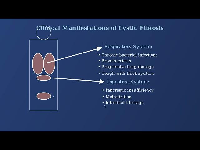
Cystic fibrosis affects multiple organ
10:46
systems throughout the body with varying
10:48
clinical manifestations. In the
10:50
respiratory system, thick mucus leads to
10:52
chronic bacterial infections,
10:54
bronchiacttois, and progressive lung
10:56
damage. The digestive system is affected
10:59
by pancreatic insufficiency leading to
11:02
malnutrition and difficulty digesting
11:04
fats and proteins. In the reproductive
11:07
system, 98% of males with cystic
11:09
fibrosis are infertile due to congenital
11:12
absence of the vas deference.
11:15
While cystic fibrosis affects multiple
11:17
organ systems, progressive lung disease
11:20
remains the primary cause of morbidity
11:22
and mortality. Advances in treatment
11:25
have dramatically increased the median
11:27
survival age from childhood to over 40
11:30
years in developed countries.
11:32
Huntington's disease is a progressive
11:34
neurodeenerative disorder that affects
11:37
the brain's nerve cells. It is
11:39
characterized by three main symptom
11:41
categories. Uncontrolled movements known
11:43
as Korea, emotional problems including
11:46
depression and irritability, and
11:48
cognitive decline affecting memory and
11:50
decision-m. The disease typically
11:52
manifests in adulthood, usually between
11:55
the ages of 30 and 50, though juvenile
11:58
onset forms exist. It has a prevalence
12:01
of approximately 5 to 10 per 100,000
12:04
people in western populations.
12:07
Huntington's disease follows an
12:09
autotosomal dominant inheritance
12:10
pattern, meaning that a single copy of
12:13
the mutated gene from either parent is
12:15
sufficient to cause the disease. The
12:18
disease is caused by a mutation in the
12:20
HTT gene, which leads to the production
12:22
of an abnormal form of the Huntington
12:24
protein that gradually damages neurons
12:27
in the brain.
12:30
Understanding the clinical features and
12:31
epidemiology of Huntington's disease is
12:34
essential for diagnosis and patient
12:36
management.
12:38
Huntington's disease is caused by a
12:39
mutation in the HTT gene located on
12:42
chromosome 4. The mutation is
12:44
characterized by an expanded KAG
12:46
trucleotide repeat in the coding region
12:48
of the gene. In healthy individuals,
12:51
this gene contains around 17 to 20
12:53
repeats of the KAG sequence. But in
12:56
Huntington's disease, this repeat is
12:58
expanded to 35 or more copies.
13:02
This expanded KAG repeat leads to the
13:04
production of an abnormal Huntington
13:06
protein with an extended stretch of
13:08
glutamine amino acids resulting in
13:10
protein misfolding and aggregation.
13:14
There is a strong inverse correlation
13:16
between the number of KAG repeats and
13:18
the age of disease onset. Individuals
13:21
with longer repeats typically develop
13:23
symptoms at an earlier age.
13:26
Huntington's disease follows an
13:28
autotosomal dominant inheritance
13:30
pattern. This means that a person only
13:33
needs to inherit one copy of the mutated
13:35
gene to develop the disease. Each child
13:38
of an affected parent has a 50% chance
13:41
of inheriting the mutation and
13:43
developing the disease. This pattern of
13:46
inheritance makes Huntington's disease
13:48
particularly devastating for families.
13:53
Molecular testing for Huntington's
13:55
disease primarily focuses on determining
13:58
the number of KAG repeats in the HTT
14:00
gene. PCRbased methods are the gold
14:03
standard for determining KAG repeat
14:05
length in the Huntington gene. The PCR
14:08
testing process involves DNA extraction,
14:11
amplification of the region containing
14:12
KAG repeats, and fragment analysis to
14:15
determine the exact number of repeats.
14:18
CAG repeat length determines disease
14:20
status. Normal individuals have 26 or
14:23
fewer repeats while full penetrance
14:25
occurs with 40 or more repeats leading
14:28
to certain disease development.
14:30
As the KAG repeat number increases,
14:32
particularly above 40 repeats, the
14:35
expanded HTT gene leads to the
14:37
production of mutant Huntington protein.
14:41
For very large CCAG expansions, southern
14:43
blotting may be used as a confirmatory
14:45
method when PCR results are ambiguous or
14:47
suggest extremely large repeat numbers.
14:51
Southern blotting involves restriction
14:52
enzyme digestion of genomic DNA, gel
14:55
electropharesis to separate fragments by
14:58
size, transfer to a membrane, and
15:00
detection using a labeled probe.
15:03
Southern blotting is particularly
15:05
indicated when PCR results suggest very
15:07
large expansions, when PCR shows
15:10
homozygosity for normal alals, or when
15:13
technical difficulties are encountered
15:14
with PCR amplification.
15:17
Testing for Huntington's disease raises
15:20
significant ethical considerations as it
15:22
is a late onset disorder with no current
15:25
cure. Ethical challenges include the
15:27
necessity for pre-est genetic
15:29
counseling, informed consent that
15:31
addresses psychological implications,
15:34
and concerns about testing asymptomatic
15:36
minors. Results may impact family
15:38
planning decisions and potentially lead
15:40
to insurance or employment
15:42
discrimination.
15:45
International testing protocols for
15:47
Huntington's disease require multiple
15:49
counseling sessions, psychological
15:51
assessment and support, confirmation of
15:54
results with a second independent
15:56
sample, and long-term follow-up with
15:58
medical and psychological support.
16:04
Huntington's disease is caused by a
16:06
mutant Huntington protein that contains
16:08
an expanded CAG repeat resulting in an
16:11
abnormally long polyglutamine tract. The
16:14
mutant protein misfolds and forms toxic
16:16
aggregates within neurons particularly
16:18
in the brain's basil ganglia and
16:20
cerebral cortex.
16:23
The accumulation of mutant huntington
16:25
primarily affects specific brain
16:27
regions. The basil ganglia particularly
16:29
the strriatam show significant atrophy.
16:32
The cerebral cortex is also affected
16:35
with progressive neuronal loss leading
16:37
to the characteristic cognitive and
16:39
psychiatric symptoms.
16:42
Huntington's disease is characterized by
16:43
a clinical triad of symptoms motor
16:46
cognitive and psychiatric
16:48
manifestations. Motor symptoms include
16:50
korea, which refers to involuntary,
16:52
irregular dance-like movements, as well
16:55
as donia and impaired coordination.
16:57
Cognitive decline manifests as impaired
17:00
memory, judgment, and problem solving
17:02
abilities, which worsen as the disease
17:04
progresses. Psychiatric symptoms often
17:07
include depression, anxiety, and
17:09
irritability, which can precede motor
17:11
symptoms by many years.
17:14
Brain imaging reveals the structural
17:16
changes in Huntington's disease. In a
17:18
normal brain, the stratum appears
17:20
healthy and ventricles are of normal
17:22
size. In contrast, the HD brain shows
17:26
significant stridal atrophy, enlarged
17:28
ventricles, and overall brain volume
17:30
loss. It's important to note that
17:32
symptoms typically appear between ages
17:34
30 to 50 and progressively worsen over
17:36
10 to 30 years.
17:40
Cickle cell anemia is caused by a
17:42
specific mutation in the HBB gene which
17:44
is located on chromosome 11.
17:47
The HBB gene contains the instructions
17:50
for making betalobin, a component of
17:52
hemoglobin. In the normal gene, at a
17:55
specific position, there's a sequence
17:57
that codes for glutamic acid at position
17:59
six of the betalobin chain. Cickle cell
18:02
anemia is caused by a single nucleotide
18:04
substitution where an adinine A is
18:07
replaced by thymine T in the DNA
18:09
sequence. This substitution changes the
18:11
codon from gag to GTG which results in
18:14
the amino acid veiling replacing
18:16
glutamic acid at position 6 of the
18:18
betalobin chain.
18:21
This mutation creates hemoglobin S HBS
18:24
instead of normal hemoglobin A HBA. The
18:27
key difference is that glutamic acid is
18:29
hydrophilic or water loving while veene
18:31
is hydrophobic or water repelling.
18:34
This single amino acid change has
18:36
profound effects on the hemoglobin's
18:38
behavior. In low oxygen conditions,
18:41
hemoglobin s molecules stick together
18:43
forming rigid polymers that distort the
18:46
red blood cell into the characteristic
18:48
sickle shape. This demonstrates how a
18:50
single nucleotide substitution can lead
18:52
to dramatic changes in protein structure
18:55
and function resulting in a serious
18:57
genetic disease.
19:00
Molecular testing for cickle cell anemia
19:02
employs several diagnostic methods to
19:05
detect the specific mutation in the
19:07
betalobin gene. These methods include
19:09
PCR RFLP analysis, alals specific PCR
19:12
DNA sequencing and hemoglobin
19:14
electropharesis for initial screening.
19:18
PCR RFLP analysis is one of the most
19:20
commonly used methods. It involves
19:22
amplifying the betalobin gene region
19:24
containing the cickle cell mutation
19:26
using PCR. The PCR product is then
19:29
digested with restriction enzymes like
19:31
DDI or MES2. The cickle cell mutation
19:35
eliminates a restriction site creating
19:36
fragments of different lengths. Gel
19:38
electropharesis separates these
19:40
fragments by size producing distinct
19:43
band patterns for normal carrier and
19:45
cickle cell genotypes.
19:48
Al specific PCR uses primers designed to
19:50
selectively amplify either the normal or
19:53
cickle cell al. The wild type primer
19:56
matches the normal sequence with an A at
19:58
the mutation site while the mutant
20:00
primer matches the cickle cell sequence
20:02
with a T. Here we can see the critical
20:04
difference in DNA sequence. In the
20:07
normal alil we have gag coding for
20:09
glutamic acid while in the cickle al we
20:12
have gtg coding for veene. Samples are
20:15
analyzed by gel electropharesis to
20:17
determine which primers produced
20:19
amplification products revealing the
20:21
patient's genotype.
20:23
DNA sequencing is the gold standard for
20:26
detecting the cickle cell mutation
20:28
providing direct visualization of the A
20:30
to T substitution in the betalobin gene.
20:33
In a sequencing chromatog, we can
20:35
clearly see the difference between a
20:37
normal sample showing gag glutamic acid
20:40
and a cickle cell sample showing GTG
20:43
veene. Sequencing can also detect rare
20:45
betagalobin mutations and is often used
20:48
for confirmatory testing after initial
20:50
screening.
20:52
Newborn screening programs use
20:54
hemoglobin electrofpharesis as an
20:56
initial test to identify infants with
20:58
cickle cell disease. This technique
21:01
separates normal hemoglobin A from
21:03
cickle hemoglobin S based on their
21:05
different electrical charges.
21:07
Isoelectric focusing provides better
21:09
resolution for distinguishing various
21:11
hemoglobin variants. Positive screening
21:14
results are confirmed with molecular
21:16
diagnostic methods like those we've
21:18
discussed. Early diagnosis through
21:21
newborn screening allows for immediate
21:23
intervention with prophylactic
21:25
antibiotics and parent education,
21:27
significantly reducing mortality.
21:32
Molecular testing methods provide the
21:34
foundation for accurate diagnosis,
21:36
carrier detection, and prenatal testing
21:38
for cickle cell anemia.
21:43
Cickle cell anemia results from a single
21:46
point mutation in the betaglobin gene.
21:49
In this mutation, a hydrophilic glutamic
21:52
acid is replaced by a hydrophobic veene
21:54
at position six of the betalobin chain.
21:57
This substitution significantly alters
21:59
the properties of hemoglobin causing it
22:01
to polymerize under low oxygen
22:03
conditions.
22:06
Let's examine how the sickling process
22:08
occurs in red blood cells. Normal red
22:10
blood cells are flexible and discshaped,
22:13
allowing them to easily navigate through
22:15
blood vessels. Under low oxygen
22:17
conditions, the mutated hemoglobin
22:19
molecules polymerize into rigid fibers.
22:22
This causes the red blood cells to lose
22:24
their flexibility and take on the
22:26
characteristic sickle or crescent shape.
22:29
Initially, this sickling process is
22:31
reversible when oxygen levels are
22:33
restored. However, with repeated cycles,
22:36
the damage becomes permanent.
22:39
Cickle cell anemia leads to several
22:41
serious clinical manifestations.
22:46
One of the primary manifestations is
22:48
hemolytic anemia. Sickled red blood
22:50
cells are rigid and fragile making them
22:53
prone to destruction. Their lifespan is
22:55
dramatically reduced from the normal 120
22:58
days to only 10 to 20 days. This chronic
23:01
hemolysis leads to persistent anemia and
23:04
jaundice due to the release of bilerubin
23:06
from destroyed cells. The body attempts
23:09
to compensate by increasing red blood
23:11
cell production leading to bone marrow
23:13
expansion.
23:16
Vasoclusive crisis is the most common
23:18
and painful complication of cickle cell
23:20
anemia. In normal circulation, red blood
23:24
cells flow freely through blood vessels.
23:26
However, sickled cells can become stuck
23:28
in small blood vessels, blocking blood
23:31
flow to tissues. This occlusion is
23:33
worsened by increased adhesion of
23:34
sickled cells to vessel walls and to
23:37
each other.
23:38
The result is severe pain known as a
23:41
pain crisis which is the hallmark
23:43
clinical feature of cickle cell disease.
23:45
These crises can affect any organ in the
23:47
body but commonly involve the bones,
23:49
chest and abdomen.
23:52
Cickle cell anemia leads to several
23:54
other serious clinical manifestations.
23:56
Acute chest syndrome is a
23:58
life-threatening complication
24:00
characterized by pulmonary vaso
24:01
acclusion and often infection. Patients
24:04
also have increased susceptibility to
24:06
infections particularly from
24:08
encapsulated bacteria. This is due to
24:11
functional esplenia as repeated splenic
24:13
infarks lead to a non-functioning
24:15
spleen. Additional complications include
24:18
stroke, pulmonary hypertension, and
24:20
chronic damage to organs including the
24:22
kidneys, liver, and heart.
24:25
Carrier testing identifies individuals
24:28
who carry one copy of a gene mutation
24:30
for a disease that requires two copies
24:32
for an individual to be affected.
24:36
Carrier testing is commonly performed
24:38
for several recessive disorders. Two key
24:40
examples we've discussed are cystic
24:42
fibrosis caused by mutations in the CFTR
24:45
gene and cickle cell anemia caused by
24:48
mutations in the hemoglobin beta gene.
24:51
To understand carrier status, we need to
24:54
look at chromosome pairs. In a normal
24:56
individual, both chromosomes have
24:58
functioning genes. A carrier has one
25:00
normal copy and one mutated copy and
25:03
typically shows no symptoms. An affected
25:06
individual has mutations in both copies
25:08
of the gene. It's important to note that
25:10
carriers typically don't show symptoms
25:12
of the disease, but they can pass the
25:14
mutation to their children.
25:17
Genetic counseling is a process that
25:19
helps individuals understand the
25:21
medical, psychological, and familial
25:24
implications of genetic contributions to
25:26
disease.
25:28
Genetic counseling is typically provided
25:30
by certified genetic counselors, medical
25:32
geneticists, or specialists with
25:35
genetics training.
25:37
Genetic counseling is recommended in
25:39
several scenarios. When there's a family
25:42
history of a genetic disorder, when
25:44
carrier testing results are positive,
25:47
when considering prenatal testing in
25:49
cases of conenuinity where parents are
25:51
related, or after abnormal newborn
25:54
screening results.
25:56
The genetic counseling process typically
25:58
involves several steps. Collecting
26:00
family medical history, conducting a
26:03
risk assessment, discussing testing
26:05
options, interpreting results, and
26:07
providing support and followup. It's
26:09
important to understand that genetic
26:11
counseling addresses both medical and
26:14
psychological aspects, helping
26:16
individuals make informed decisions
26:18
about testing and family planning.
26:21
A pedigree is a chart that represents
26:24
family relationships and the inheritance
26:26
pattern of genetic traits across
26:28
generations.
26:30
Pedigrees use standardized symbols to
26:32
represent individuals and their genetic
26:34
status. Males are represented by
26:37
squares, females by circles. Filled
26:39
symbols indicate affected individuals,
26:42
while a dot inside a symbol indicates a
26:44
carrier of a recessive disorder.
26:47
Let's examine different inheritance
26:49
patterns in pedigrees. Here's an
26:51
autosomal recessive pattern seen in
26:53
diseases like cystic fibrosis and cickle
26:56
cell anemia. In this pattern, carriers
26:59
shown with dots have one mutation but
27:01
aren't affected, while individuals with
27:03
two mutations show symptoms. In
27:06
autotosomal dominant inheritance, as
27:08
seen in Huntington's disease, having
27:10
just one copy of the mutated gene is
27:12
enough to cause disease. Each child of
27:15
an affected parent has a 50% chance of
27:17
inheriting the mutation. X- linked
27:20
recessive disorders like hemophilia show
27:23
a distinct pattern where males are more
27:25
commonly affected than females. Carrier
27:28
females with one mutation can pass the
27:30
condition to their sons who may be
27:32
affected or to their daughters who may
27:34
become carriers. Pedigree analysis helps
27:37
determine the pattern of inheritance
27:39
which is crucial for accurate risk
27:41
assessment in genetic counseling.
27:44
Risk calculations determine the
27:46
probability of passing a genetic
27:48
condition to offspring based on
27:50
inheritance patterns.
27:53
For autosomal recessive conditions like
27:54
cystic fibrosis when both parents are
27:57
carriers having one normal gene A and
27:59
one mutated gene A, we can use a Punet
28:02
square to calculate risk. Each child has
28:04
a 25% chance of inheriting two mutated
28:07
copies and being affected.
28:10
For autotosomal dominant conditions like
28:12
Huntington's disease, when one parent is
28:14
affected, having one copy of the
28:16
dominant mutated gene D, each child has
28:19
a 50% chance of inheriting the mutation
28:22
and developing the disease.
28:25
X- linked recessive conditions like
28:27
hemophilia show different inheritance
28:29
patterns. When a mother is a carrier and
28:32
the father is unaffected, 50% of sons
28:35
may be affected because they inherit
28:37
only one X chromosome. 50% of daughters
28:40
may be carriers, but they rarely show
28:42
symptoms because they have a second
28:44
normal X chromosome.
28:47
Understanding carrier status and risk
28:49
calculations is crucial in genetic
28:51
counseling. It provides accurate risk
28:54
assessment for informed family planning,
28:57
enables early intervention for
28:58
potentially affected children, helps
29:01
with psychological preparation, and
29:03
informs decisions about further testing
29:05
options. Ultimately, carrier testing and
29:08
genetic counseling empower individuals
29:11
to make informed decisions about their
29:13
genetic health and family planning.
29:17
Prenatal genetic testing allows for the
29:19
detection of genetic abnormalities
29:21
before birth. There are several methods
29:24
available, each with different timing,
29:26
accuracy, and risk profiles.
29:29
Amnocentesis involves sampling amniotic
29:32
fluids surrounding the fetus. This
29:34
procedure is typically performed between
29:36
15 and 20 weeks of pregnancy and
29:39
provides highly accurate diagnostic
29:41
information but carries a small risk of
29:44
miscarriage. Corionic villa sampling or
29:46
CVS samples tissue from the placenta. It
29:49
can be performed earlier than
29:51
amnocentesis typically between 10 and 13
29:54
weeks. While also highly accurate, it
29:56
has a slightly higher miscarriage risk.
29:59
Non-invasive prenatal testing analyzes
30:01
cell-free fetal DNA circulating in the
30:04
mother's bloodstream. This screening
30:06
test can be performed as early as 10
30:08
weeks and poses no risk to the
30:10
pregnancy, but it's not as definitive as
30:12
diagnostic tests like amnocentesis or
30:15
CVS.
30:17
These prenatal tests can detect a wide
30:19
range of genetic conditions including
30:21
chromosomal abnormalities like Down
30:24
syndrome, single gene disorders like
30:26
cystic fibrosis, neural tube defects,
30:29
and various inherited genetic disorders.
30:33
Pre-implantation genetic testing or PGT
30:36
is performed on embryos created through
30:38
invitro fertilization before
30:40
implantation in the uterus. The process
30:43
begins with in vitro fertilization where
30:45
eggs are harvested and fertilized with
30:47
sperm in a laboratory setting. These
30:50
fertilized eggs develop into early
30:52
embryos typically reaching the blast
30:54
stage after 5 to 6 days. At this point,
30:57
a biopsy is performed where a few cells
31:00
are carefully removed from each embryo
31:02
for genetic testing. These cells undergo
31:05
genetic analysis to detect specific
31:07
genetic conditions or chromosomal
31:09
abnormalities that the parents may be at
31:12
risk of passing on. Based on the
31:14
results, embryos without detected
31:16
genetic abnormalities are selected for
31:18
transfer to the woman's uterus,
31:20
increasing the chances of a healthy
31:22
pregnancy.
31:24
There are several types of
31:25
pre-implantation genetic testing. PGTA
31:28
tests for anuploloy or abnormal
31:30
chromosome numbers. PGTM tests for
31:33
specific monogenic or single gene
31:35
disorders and PGTSR tests for structural
31:38
rearrangements like chromosomeal
31:39
transllocations.
31:43
Both prenatal and pre-implantation
31:45
genetic testing raise important ethical
31:47
considerations that patients, healthcare
31:49
providers, and society must navigate.
31:52
From a medical perspective, it's
31:54
important to consider the accuracy and
31:56
limitations of these tests, the
31:59
potential risks of invasive procedures
32:01
to the mother or fetus, and the
32:03
possibility of incidental findings that
32:05
weren't specifically being tested for.
32:08
There are also broader social and
32:09
personal considerations, including
32:12
reproductive autonomy, disability rights
32:14
perspectives, and concerns about the
32:16
potential for genetic selection based on
32:18
non-medical traits. The importance of
32:21
informed consent and genetic counseling
32:23
cannot be overstated. These services
32:26
help patients understand the
32:28
implications of testing and make
32:30
informed decisions aligned with their
32:32
personal values.
32:36
Molecular diagnostics continues to
32:38
evolve with several emerging
32:39
technologies transforming genetic
32:41
testing capabilities. Three key
32:43
technologies at the forefront include
32:45
crisper-based diagnostics, digital PCR,
32:49
and single cell sequencing.
32:51
Crisper-based diagnostic systems like
32:53
Sherlock and detector leverage the
32:55
precise targeting capabilities of
32:57
crisper cast proteins. These systems can
33:00
identify specific nucleic acid sequences
33:02
with singlebased precision.
33:05
These diagnostics offer several
33:06
advantages. high specificity,
33:09
isoothermal detection without requiring
33:11
complex equipment, rapid results within
33:13
minutes, and adaptability to portable
33:16
testing formats for use in resource
33:18
limited settings.
33:20
Digital PCR represents a significant
33:22
advancement over traditional PCR
33:24
methods. It partitions the sample into
33:26
thousands of nanolized droplets or
33:29
chambers with each partition acting as
33:31
an individual PCR reaction. This
33:34
partitioning enables absolute
33:36
quantification without standard curves,
33:38
higher precision for detecting low
33:40
abundance targets, enhanced sensitivity
33:42
for rare mutations, and better tolerance
33:45
to inhibitors that might affect
33:47
traditional PCR.
33:50
Single cell sequencing technologies
33:52
allow us to analyze the genetic material
33:54
from individual cells rather than bulk
33:56
tissue samples. This provides
33:58
unprecedented resolution of cellular
34:01
differences within seemingly homogeneous
34:03
tissues. The applications are
34:05
transformative, revealing cellular
34:07
heterogeneity in tissues, identifying
34:10
rare cell populations that may be
34:12
disease driving, tracking clonal
34:14
evolution in cancer, and mapping
34:16
developmental trajectories in complex
34:18
biological systems.
34:21
A major advancement is the adaptation of
34:23
these technologies for point of care
34:25
testing, moving molecular diagnostics
34:28
from centralized laboratories to patient
34:31
bedsides and field settings. The
34:33
benefits include rapid results where
34:35
patients are located, expanded testing
34:37
capacity in resource limited settings,
34:39
real-time clinical decisionmaking, and
34:42
significant reduction in time from
34:44
testing to treatment. Importantly,
34:47
these technologies are advancing toward
34:49
complete sample to answer workflows that
34:52
require minimal technical expertise,
34:55
making them ideal for decentralized
34:57
testing environments.
35:00
These emerging technologies are
35:02
transforming molecular diagnostics
35:04
through significant improvements in test
35:06
sensitivity, specificity, and
35:08
accessibility. Crisper-based systems
35:10
offer exceptional specificity and
35:13
growing accessibility. Digital PCR
35:15
provides unmatched sensitivity for rare
35:18
targets. Single cell sequencing delivers
35:20
unprecedented resolution of cellular
35:22
heterogeneity.
35:25
Gene therapy approaches use genetic
35:27
material to treat or prevent diseases
35:29
caused by genetic mutations.
35:33
Molecular diagnostics is crucial for
35:35
gene therapy as it identifies specific
35:37
mutations, determines therapeutic
35:39
targets, guides vector selection, and
35:42
measures treatment efficacy.
35:45
For cystic fibrosis, viral vector
35:47
approaches use adnoassociated viruses
35:49
and lentiviruses to deliver functional
35:51
CFTR genes to airway epithelial cells.
35:54
Recent clinical trials have shown
35:56
promising results with improved lung
35:58
function and reduced infection frequency
36:00
in patients.
36:03
For cickle cell anemia, crisper cast 9
36:06
gene editing approaches directly correct
36:08
the mutation in the betalobin gene or
36:10
induce fetal hemoglobin expression. In a
36:13
major breakthrough, the FDA approved
36:15
CASG in 2023, the first crisper-based
36:19
therapy for cickle cell anemia. For
36:21
Huntington's disease, RNA interference
36:23
strategies use anti-sense olonucleotides
36:26
and small interfering RNAs to silence
36:29
the expression of the mutant Huntington
36:31
gene. Multiple clinical trials are
36:34
currently underway with promising safety
36:36
profiles and early indications of
36:38
reduced mutant huntington protein
36:40
levels.
36:42
Each genetic disease requires a
36:44
different gene therapy approach with
36:46
specific delivery methods tailored to
36:48
the affected tissues. Gene therapy
36:51
approaches continue to advance with
36:53
improved delivery systems, reduced
36:55
immune responses, and enhanced targeting
36:57
specificity.
37:00
Pharmaccogenomics enables personalized
37:02
medicine by tailoring treatments based
37:04
on a patient's unique genetic profile.
37:07
Pharmaccogenomics studies how genes
37:09
affect a person's response to drugs,
37:11
enabling more effective and safer
37:13
treatment approaches.
37:16
The personalized medicine approach
37:18
begins with genetic testing to identify
37:21
variants that affect drug response.
37:23
Next, clinicians analyze these genetic
37:26
results to determine which medications
37:28
will be most effective for the patient.
37:30
Finally, this information guides
37:32
personalized treatment decisions,
37:34
including drug selection, dosing, and
37:36
monitoring.
37:39
Let's look at disease specific
37:40
applications of pharmaccogenomics. In
37:43
cystic fibrosis, CFTR modulators are
37:45
designed to target specific mutations.
37:48
Ivactor works for patients with G551D
37:51
and other gating mutations. Lumacater
37:53
combined with ivaca known as orcami was
37:56
developed specifically for patients
37:57
homozygous for the F508 Dell mutation.
38:01
The triple combination therapy trafta
38:03
expanded treatment options for patients
38:05
with at least one F508 Dell mutation
38:08
benefiting about 90% of CF patients.
38:13
For other genetic disorders, targeted
38:15
therapies are also emerging. In
38:16
Huntington's disease, anti-sense
38:18
oligouonucleotides target the mutant HTT
38:21
gene and RNA interference therapies aim
38:24
to reduce toxic protein production. For
38:26
cickle cell anemia, hydroxyura increases
38:29
fetal hemoglobin production. Gene
38:31
therapy approaches targeting the HBB
38:33
gene and crisper gene editing therapies
38:36
are also in development.
38:39
Personalized medicine offers several key
38:42
benefits, including improved efficacy,
38:44
and reduce side effects from treatments.
38:47
It reduces trial and error prescribing,
38:49
enables better prediction of disease
38:51
progression, optimizes drug dosing, and
38:54
helps prevent adverse drug reactions.
38:56
However, challenges remain, including
38:58
cost and accessibility issues, clinical
39:01
implementation of genetic information,
39:03
and the need for physician education.
39:06
Despite these challenges,
39:08
pharmaccogenomic testing is increasingly
39:10
becoming standard of care for many
39:12
conditions.
39:17
Genetic testing brings numerous
39:19
benefits, but also raises significant
39:21
ethical considerations that must be
39:23
addressed. Privacy concerns are
39:26
paramount in genetic testing. Genetic
39:28
data contains highly sensitive personal
39:30
information that could potentially be
39:32
accessed by unauthorized parties. There
39:35
are ongoing questions about data
39:37
ownership, control, and the long-term
39:40
implications of storing genetic
39:41
information.
39:44
Genetic testing results could
39:46
potentially lead to discrimination in
39:48
employment or insurance coverage based
39:50
on genetic predispositions to disease.
39:53
While laws like the Genetic Information
39:55
Non-Discrimination Act provide some
39:58
protections, they have limitations and
40:00
don't cover all scenarios.
40:04
Genetic tests may reveal incidental
40:07
findings, genetic variance unrelated to
40:10
the original purpose of testing. These
40:12
can include variance of uncertain
40:14
significance or predispositions to other
40:17
conditions, raising questions about the
40:19
responsibility to disclose and the
40:21
patients right not to know certain
40:23
information.
40:26
The psychological impact of genetic test
40:28
results can be profound. Positive
40:31
results may cause anxiety and distress,
40:34
while negative results can lead to
40:35
survivor guilt when other family members
40:37
are affected. Test results can change a
40:40
person's self-perception and identity
40:42
and significantly impact family dynamics
40:45
and relationships.
40:49
Informed consent is essential in genetic
40:51
testing. This includes full disclosure
40:54
of the testing purpose and limitations,
40:56
explanation of possible results and
40:58
their implications, and discussion of
41:00
data storage and sharing policies.
41:03
Special considerations are needed for
41:05
vulnerable populations, including
41:07
children and those with cognitive
41:08
impairments.
41:12
Genetic counseling plays a crucial role
41:15
both before and after testing. Pre-EST
41:18
counseling ensures informed
41:19
decision-making while post- test
41:21
counseling helps explain results and
41:23
guide next steps. Counselors provide
41:25
vital support for the emotional and
41:27
psychological impacts of genetic test
41:29
results.
41:32
Ultimately, genetic testing requires
41:34
careful balancing of benefits and risks.
41:37
Ethical frameworks help navigate these
41:39
complex considerations, ensuring that
41:42
genetic testing serves the patients best
41:44
interests while respecting their
41:46
autonomy and dignity.
41:51
We now summarize the key points about
41:53
molecular diagnosis of genetic diseases.
41:56
For cystic fibrosis, molecular diagnosis
41:59
centers on identifying mutations in the
42:01
CFTR gene with F508 Dell being the most
42:04
common. Various techniques including
42:06
RFLP, next generation sequencing, and
42:08
MLPA are employed for accurate
42:10
detection. In Huntington's disease,
42:12
diagnosis focuses on the KAG repeat
42:14
expansion in the HTT gene. This
42:17
molecular understanding explains the
42:19
anticipation phenomenon where symptoms
42:21
appear earlier in successive
42:23
generations. Cickle cell anemia involves
42:26
a single point mutation in the HBB gene
42:29
leading to abnormal hemoglobin S
42:31
production and characteristic
42:33
sickle-shaped red blood cells. Newborn
42:36
screening has been crucial in preventing
42:37
complications. The clinical impact of
42:40
molecular diagnostics has been
42:42
transformative across these conditions.
42:45
Early detection now enables proactive
42:47
management of symptoms and
42:48
complications.
42:50
Carrier screening has significantly
42:52
reduced disease transmission.
42:55
Targeted therapies directly address the
42:57
molecular defects and predictive testing
43:00
empowers patients to make informed
43:01
decisions about their health and family
43:03
planning. The broader public health
43:05
impact of these advances has been
43:07
substantial. Early detection has reduced
43:09
both morbidity and mortality. Carrier
43:12
screening programs have decreased
43:14
disease incidents and health care costs.
43:17
Targeted therapies have improved quality
43:19
of life and increased life expectancy
43:22
for affected individuals. Looking toward
43:24
the future, molecular diagnostics
43:26
continues to revolutionize disease
43:28
management, enabling personalized
43:30
treatment approaches and improving
43:32
patient outcomes across a wide range of
43:34
genetic conditions. As our understanding
43:37
of molecular mechanisms continues to
43:39
grow, so too will our ability to
43:42
diagnose, prevent, and treat genetic
43:44
diseases.
Genetic Disorders
Jobs & Education