Up next in 10

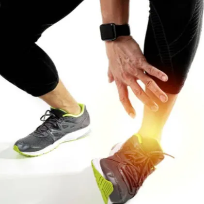
One of the more common causes of pain on the inside of the foot is due to irritation of an extra bone known as an accessory navicular bone.
Read the complete article at https://www.foot-pain-explained.com/accessory_navicular.html
@marcbarry1000
Show More Show Less View Video Transcript
0:00
OS Tibi
0:01
navicular also known as OS Tibi
0:04
navicular OS Tibi
0:07
externum what is an accessory
0:09
navicular an accessory bone is a bone
0:12
that is not normally found in the
0:14
average human but in most cases is not
0:16
considered abnormal OS Tibi navicular
0:20
refers to an extra bone found in the
0:22
foot this condition represents a
0:25
secondary oifc Center growth center of
0:28
the navicular bone
0:30
it is present from birth the navicular
0:33
bone is found on the inside part of the
0:35
foot this condition has been observed in
0:38
multiple family members and has
0:40
propensity to occur slightly more in
0:42
females than males for most individuals
0:45
this condition is a non-issue and most
0:47
people who have them never have a
0:49
problem with them in an ideal situation
0:52
the navicular bone and the accessory
0:54
bone will fuse together to form one bone
0:58
the problem that occurs is that some
1:00
times the two bones do not fuse together
1:02
and the patient is left with what is
1:04
known as a fibrous Union or basically a
1:06
non-solid union of bone to bone this
1:09
fibrous Union is more like scar tissue
1:12
and in theory can cause pain when
1:14
excessive strain is placed upon it
1:17
adding to the drama is the fact that the
1:19
accessory bone resides in the posterior
1:21
tibial tendon this tendon by Design
1:25
helps lift the arch of the foot so as
1:27
you might expect there is a tremendous
1:29
amount of tension on this tendon when a
1:31
person
1:32
ambulates people with excessively flat
1:35
feet tend to put more tension on this
1:37
tendon and thus may be more prone to
1:39
suffering pain from an accessory
1:41
navicular what causes accessory
1:43
navicular pain accessory navicular
1:46
syndrome as it is called can result from
1:49
a number of
1:50
causes excess or overuse syndrome as
1:53
seen in an athlete trauma to the foot as
1:56
in an ankle sprain or direct trauma to
1:58
the navicular bone chronic irritation
2:01
from shoes rubbing against the extra
2:03
bone over time may cause pain excessive
2:07
pronation which strains the attachment
2:09
of tibialis posterior muscles into the
2:11
navicular bone keep in mind the larger
2:15
the actual accessory bone the greater
2:17
the chance of it becoming an issue
2:20
symptoms of accessory navicular pain
2:23
symptoms of this syndrome would include
2:25
redness swelling and tenderness over the
2:27
navicular bone the I bone is located on
2:31
the inside of the foot approximately
2:33
midway between the ankle bone and big
2:35
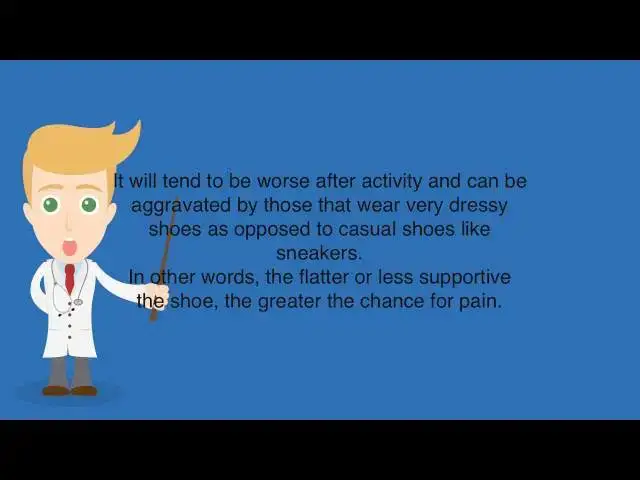
toe joint it will tend to be worse after
2:38
activity and can be aggravated by those
2:41
that wear very dressy shoes as opposed
2:43
to casual shoes like
2:45
sneakers in other words the flatter or
2:47
less supportive the shoe the greater the
2:49
chance for pain diagnosis of an
2:53
accessory
2:54
navicular diagnosis is fairly simple
2:56
based on an examination by your doctor
3:00
he or she will palpate the navicular
3:01
bone and based on the location of pain
3:04
will suspect an accessory
3:06
navicular the doctor will also observe
3:09
your gate to see if you are flat foot at
3:12
this point an x-ray will make the
3:13
definitive
3:15
diagnosis other causes of pain in the
3:17
same area of the foot would include a
3:19
fracture of the navicular bone or
3:21
possibly tendinitis or even a partial
3:24
tier of the tibialis posterior tendon
3:26
that inserts into the
3:28
navicular in the cases there is usually
3:31
a history of trauma people with a
3:34
naturally large navicular bone may also
3:36
develop atis due to Chronic shoe
3:39
pressure accessory navicular treatment
3:42
treatment is broken down into
3:44
conservative versus surgical care most
3:47
cases of accessory navicular syndrome
3:49
may be treated conservatively with some
3:52
sort of
3:54
immobilization this should allow the
3:56
fibrous tissue between the two bones to
3:58
heal if patient is extremely flat-footed
4:02
pronated then I lean more towards an
4:04
orthotic than a boot as my main goal is
4:06
to keep the patient foot from flattening
4:08
out too much and thus reduce the strain
4:10
on the two bones
4:12
additionally supplementation with ice
4:15
and oral anti-inflammatory
4:18
medication if the patient is athletic
4:21
sometimes we can keep them active with
4:23
an orthotic but other times they have to
4:25
give up their sport for a period of time
4:27
to allow the condition to heal
4:30
if conservative care does not alleviate
4:32
the problem then surgical intervention
4:34
should be
4:35
considered the most common procedure for
4:38
this condition is known as the kidner
4:40
procedure where a small incision is made
4:42
over the navicular bone the accessory
4:45
navicular is identified and dissected
4:47
free from the posterior tibial tendon
4:50
the posterior tibial tendon is then
4:52
reattached to the remaining navicular
4:54
bone read the complete article at www
5:00
f o t- p a i n- x p l a i n
5:04
d.com C SS o r ycore n a v i c u l a r
5:10
HTML
Pain Management
Other
Injury