Up next in 10

Understanding Schistosoma haematobium
Website: https://biologynotesonline.com/
Facebook: https://www.facebook.com/biologynotesonline
Instagram: https://www.instagram.com/biologynotesonline/?hl=en
Show More Show Less View Video Transcript
0:00
Hey everyone. Today we're diving into
0:02
the world of parasites, specifically
0:04
schistosoma hematobium.
0:07
This fascinating yet troublesome
0:09
organism is something every student of
0:11
biology and medicine should understand.
0:13
Schistasoma hematobium is what we call a
0:15
blood fluke. It's a type of parasitic
0:18
worm that belongs to a group called
0:20
tremodess. These are flatworms that have
0:23
adapted to live inside other organisms.
0:25
Here we can see the actual structure of
0:27
this parasite under a microscope.
0:30
Notice the oral sucker at the front
0:32
which it uses to attach to blood vessels
0:34
and the acetabulum, another sucker that
0:37
helps it stay in place. This blood fluke
0:39
causes a disease called eurogenital
0:41
schistosomiis.
0:43
The name tells us exactly what it
0:45
affects the eurogenital system which
0:48
includes the bladder, kidneys, and
0:50
reproductive organs. Think of shistasoma
0:52
hematobium as a tiny unwelcome guest
0:55
that moves into your body and refuses to
0:57
leave. Unlike a good guest, this
1:00
parasite doesn't just stay quietly. It
1:02
actively damages the places where it
1:04
lives. The key takeaway here is that
1:06
schistosoma hematobium is not just any
1:08
parasite. It's specifically adapted to
1:11
live in human blood vessels and cause
1:13
problems in our urinary and reproductive
1:16
systems. Understanding what it is helps
1:18
us better understand how to fight it.
1:21
Schistosoma hematobium has a very
1:24
specific geographic distribution around
1:26
the world. This parasite is primarily
1:28
found in two main regions. The first and
1:31
most important region is Africa where
1:33
the vast majority of infections occur.
1:36
Nearly every country in subsaharan
1:38
Africa reports cases of this parasite.
1:40
The second region is the Middle East,
1:43
particularly countries around the
1:44
eastern Mediterranean and parts of the
1:46
Arabian Peninsula. Now, let's understand
1:49
why this parasite thrives in these
1:51
specific regions. The key factor is the
1:54
presence of freshwater environments.
1:56
Schistosoma hematobium requires
1:58
freshwater environments to complete its
2:00
life cycle. This includes natural bodies
2:03
of water like lakes and rivers. It also
2:05
thrives in human-made water systems,
2:08
particularly irrigation canals and
2:10
agricultural water sources where people
2:12
frequently come into contact with water.
2:15
These freshwater environments are
2:16
crucial because they support the snail
2:18
species that serve as intermediate hosts
2:20
for the parasite. Here we can see
2:22
various snail species and microscopic
2:25
organisms. The parasite depends on
2:27
specific freshwater snail species to
2:29
complete part of its life cycle, which
2:32
is why it's only found in regions where
2:33
these snails can survive.
2:36
The geographic distribution of this
2:38
parasite has enormous public health
2:40
implications.
2:42
Over 250 million people worldwide are
2:44
currently affected by cystosomiasis
2:47
with shistoma hematobium being one of
2:49
the major species responsible. Even more
2:52
concerning, an estimated 779 million
2:55
people are at risk of infection,
2:57
primarily in Africa and parts of the
2:59
Middle East where the parasite is
3:01
endemic. Understanding where this
3:03
parasite is found is crucial for public
3:05
health planning, prevention strategies,
3:08
and helping travelers know which regions
3:10
require extra precautions around
3:12
freshwater contact.
3:14
Now that we know where these parasites
3:16
are found, let's take a closer look at
3:18
the adult worms themselves.
3:20
Understanding their size and exact
3:22
location in the body helps us grasp how
3:25
they cause disease. Adult schistosoma
3:28
hematobium worms are surprisingly small.
3:31
They measure between 6 to 20 mm in
3:33
length. To put this in perspective,
3:35
that's smaller than your fingernail.
3:37
Here's what these adult worms actually
3:39
look like under a microscope. Notice
3:41
their elongated curved shape and the
3:44
internal structures visible through
3:46
their translucent bodies. Now, where
3:49
exactly do these worms live in the human
3:51
body? They don't just float around.
3:54
Adult schistosoma hematobium worms have
3:57
a very specific home, the blood vessels
4:00
surrounding the urinary bladder. This
4:01
medical diagram shows the blood supply
4:04
to the urinary bladder. The adult worms
4:06
live specifically in these blood
4:08
vessels, the venus plexuses that
4:10
surround the bladder. This strategic
4:12
location allows them to release their
4:14
eggs directly into the bladder. From
4:17
this location in the blood vessels, the
4:19
adult worms can easily release their
4:21
eggs. The eggs then pass through the
4:23
bladder wall and exit the body in urine.
4:26
This is how the parasite continues its
4:28
life cycle and spreads to new hosts. Key
4:31
takeaways. Adult schistoma hematobium
4:34
worms are small measuring 6 to 20 mm.
4:37
They live in blood vessels around the
4:39
urinary bladder where they release eggs
4:41
that pass into urine. This specific
4:44
location is crucial for understanding
4:46
how the disease spreads and causes
4:48
symptoms.
4:50
The eggs of schistosoma hematobium are
4:52
the real troublemakers in this disease.
4:55
While the adult worms live quietly in
4:57
blood vessels, it's their eggs that
4:59
cause most of the problems patients
5:00
experience.
5:02
These eggs have a distinctive terminal
5:04
spine that helps identify them under a
5:07
microscope. But more importantly,
5:09
they're designed to pass through the
5:11
bladder wall and exit the body through
5:14
urine. Here's where the trouble begins.
5:16
As eggs pass through the bladder wall,
5:18
they cause significant damage. The sharp
5:21
spine and the egg itself create
5:23
inflammation and tissue damage. Multiple
5:25
eggs attempt to pass through the bladder
5:27
wall simultaneously. Each egg creates
5:30
microscopic tears and triggers an
5:32
inflammatory response from the body's
5:34
immune system.
5:36
The damaged bladder tissue can be seen
5:38
under a microscope. The inflammation
5:40
shows up as changes in cell structure
5:43
and increased immune cell activity. This
5:45
egg induced damage leads to the
5:47
characteristic symptoms of
5:48
schistosomiasis.
5:50
The most obvious sign is blood in the
5:52
urine called hematia which occurs
5:55
because the eggs damage blood vessels in
5:57
the bladder wall. Remember the eggs
6:00
serve a dual purpose. They cause disease
6:02
symptoms in humans, but they also ensure
6:05
the parasites survival by continuing the
6:08
life cycle when they're released in
6:10
urine into freshwater environments. Now,
6:12
here's where the story of schistosoma
6:14
hematobium gets really interesting. This
6:17
parasite cannot complete its life cycle
6:19
without a very specific partner,
6:21
freshwater snails. But what exactly is
6:24
an intermediate host? An intermediate
6:27
host is a living organism that harbors a
6:29
parasite during part of its development.
6:32
The parasite underos important changes
6:34
inside this host making it essential for
6:36
completing the life cycle. For cysoma
6:39
hematobium these intermediate hosts are
6:42
freshwater snails specifically those
6:44
from the bulinus genus. Here we can see
6:46
three important snail species that serve
6:48
as intermediate hosts. Bulinus forcali
6:51
and bulinus globosis are the primary
6:54
hosts for shistosoma hematobium. These
6:56
snails live in freshwater environments
6:58
like lakes, slowmoving rivers,
7:00
irrigation canals, and ponds. Now let's
7:03
understand what happens inside these
7:05
snails. The parasite doesn't just hide
7:08
in the snail. It underos crucial
7:10
developmental changes. The development
7:12
process inside the snail is fascinating.
7:15
First, a miridium penetrates the snail's
7:18
tissue. It then develops into a
7:20
sporoscyst which acts like a factory
7:22
producing many circaria. Finally, these
7:25
ccaria are released back into the water
7:27
ready to infect humans. This is why
7:29
snails are so critical to the parasite
7:32
survival. Without these specific
7:34
freshwater snails, schistoma hematobium
7:37
cannot complete its life cycle and
7:39
cannot infect humans. Understanding this
7:42
relationship is key to controlling the
7:44
disease. Now we begin exploring the
7:46
fascinating life cycle of schistosoma
7:48
hematobium. This complex cycle has
7:51
multiple steps and it all starts with a
7:53
critical first step. Eggs being released
7:56
in the urine of an infected person.
7:58
Inside an infected person, adult
8:01
schistoma hematobium worms live in the
8:03
blood vessels around the bladder. These
8:06
paired worms continuously produce eggs
8:08
as part of their reproductive cycle.
8:11
The eggs produced by these adult worms
8:13
have a distinctive appearance. They are
8:15
ovalshaped with a characteristic pointed
8:18
end called a terminal spine. Each egg
8:20
contains a developing larvae called a
8:22
miracidium.
8:25
Here's how the eggs get into the urine.
8:27
The adult worms living in the blood
8:29
vessels around the bladder continuously
8:31
release eggs. These eggs then pass
8:34
through the bladder wall and enter the
8:36
urine inside the bladder.
8:39
The critical moment comes when an
8:40
infected person urinates near or into
8:43
freshwater sources. The urine contains
8:46
thousands of eggs. And when this
8:47
contaminated urine enters lakes, rivers,
8:50
or ponds, it sets the stage for the
8:52
parasites life cycle to continue.
8:56
This diagram shows the complete life
8:58
cycle with our focus on step one
9:00
highlighted. Remember, without this
9:03
initial contamination step, eggs being
9:05
released in urine that reaches fresh
9:07
water, the entire cycle would be broken.
9:10
This is why proper sanitation and
9:12
avoiding urination near water sources is
9:14
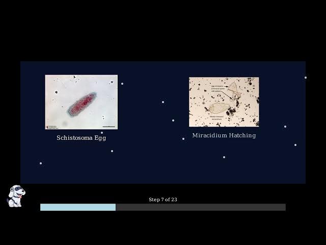
so important for prevention. When
9:16
schisto hematobium eggs reach fresh
9:19
water, something remarkable happens. The
9:21
eggs begin to hatch, releasing tiny
9:24
larve called miracy. Here we can see a
9:26
schistooma hematobium egg under the
9:28
microscope. Notice its distinctive oval
9:31
shape with the characteristic terminal
9:33
spine that helps identify this species.
9:35
When the egg comes into contact with
9:37
fresh water, it begins to hatch. The
9:39
miridium larvae emerges, leaving behind
9:42
the empty eggshell with its distinctive
9:44
terminal spine still visible.
9:47
The miracidium is a fascinating
9:49
microscopic organism. It's covered in
9:52
tiny hairike structures called psyia
9:54
that beat rapidly to propel it through
9:56
the water like a living torpedo. The
9:58
miracidium has several key features that
10:01
help it survive and find its next host.
10:03
It has eye spots that can detect light
10:05
and shadow, helping it navigate toward
10:08
potential snail hosts. It also contains
10:11
special penetration glands that will
10:13
help it burrow into a snail when it
10:14
finds one. Unlike passive organisms,
10:18
miracia are active hunters. They swim
10:20
vigorously through the water, constantly
10:23
searching for their specific snail
10:24
hosts.
10:26
Time is critical. They only have about 8
10:28
to 12 hours to find a suitable snail
10:31
before they die. Watch how the Miracidia
10:33
move through the water with purpose.
10:35
Their psyia beat in coordinated waves,
10:38
creating a spiral swimming motion that
10:40
helps them cover more area as they
10:42
search for snails. The key point to
10:45
remember is that Miracidia represent a
10:47
critical and time-sensitive stage in the
10:49
parasites life cycle. These tiny
10:52
swimming missiles must successfully
10:54
locate and penetrate a compatible snail
10:56
host within hours or the entire
10:59
reproductive cycle fails. Now we reach a
11:01
critical step in the parasites life
11:03
cycle. The miracia must find and
11:06
successfully infect their snail hosts to
11:08
continue their development. The
11:10
miracidium larvae covered in psyia for
11:12
swimming actively searches for its snail
11:15
host. These microscopic parasites can
11:17
detect chemical signals released by
11:19
snails in the water. The target is a
11:22
freshwater snail. The miridium uses
11:25
chemotaxis following chemical gradients
11:27
to locate the snail. Once found, it must
11:30
penetrate the snail's soft tissues.
11:33
The infection process happens in three
11:35
key steps. First, the miridium attaches
11:38
to the snail's surface using specialized
11:40
structures. Next, it releases powerful
11:43
enzymes that break down the snail's
11:44
tissues, creating a pathway for entry.
11:47
Finally, the miridium burrows completely
11:50
into the snail's body cavity, where it
11:52
will begin its transformation.
11:54
Once inside the snail, the miridium
11:57
underos remarkable transformations. It
12:00
first develops into a structure called a
12:01
sporosyst, then into ria. The most
12:04
important aspect is asexual
12:06
multiplication. From a single miridium,
12:09
thousands of new parasites can be
12:10
produced inside one snail host.
12:14
This snail infection step is absolutely
12:16
crucial for the parasite survival.
12:19
Without successful snail infection, the
12:21
entire life cycle breaks down. The snail
12:24
stage amplifies parasite numbers
12:25
dramatically, prepares the next
12:27
infectious stage and ensures successful
12:30
transmission back to humans. This is why
12:33
controlling snail populations is a key
12:35
strategy in preventing schistosomis.
12:38
Remember this key takeaway. The snail
12:40
infection stage is where massive
12:42
multiplication occurs. Transforming a
12:44
single miridium into thousands of
12:46
parasites ready for the next stage of
12:49
the life cycle. Inside the infected
12:51
snail, an important transformation is
12:54
taking place. The miridyia that entered
12:56
the snail are now developing into the
12:59
next stage of the parasites life cycle.
13:01
The miracia undergo a complex
13:03
transformation process inside the
13:05
snail's tissues. They develop into
13:07
circaria which are forktailed laral
13:10
forms of the parasite. This
13:12
transformation takes several weeks to
13:14
complete. Once the circaria are fully
13:16
developed, they are released from the
13:18
snail into the surrounding water. These
13:21
forktailed larve are excellent swimmers
13:23
and actively seek out their next host,
13:26
humans. Circaria have several key
13:29
characteristics that make them effective
13:30
at finding and infecting humans. They
13:33
have distinctive fork-shaped tails that
13:36
help them swim. They can survive in
13:38
fresh water for several hours, and they
13:40
actively seek out human skin to
13:42
penetrate. This release of circaria into
13:45
fresh water represents a critical point
13:48
in the parasites life cycle. The
13:50
circaria are now ready to complete their
13:52
journey by finding and infecting human
13:55
hosts who come into contact with
13:57
contaminated water. Now we reach the
13:59
critical moment when the parasite
14:01
infects humans. The circa that were
14:04
released from snails are now swimming
14:06
freely in the water actively seeking
14:08
their next host. These microscopic circa
14:12
are equipped with specialized enzymes
14:14
that allow them to penetrate human skin.
14:17
They actively swim toward any human who
14:19
enters the water. Human infection
14:22
typically occurs during everyday water
14:24
activities. People washing clothes,
14:27
children playing, or anyone bathing in
14:29
contaminated water can be exposed to
14:31
circaria. Even brief contact with the
14:34
water is enough for infection to occur.
14:37
When a circaria contacts human skin, it
14:39
uses specialized enzymes to dissolve the
14:42
outer skin barrier. The parasite then
14:44
penetrates through the skin layers
14:46
leaving behind its tail as it burrows
14:48
deeper into the tissue.
14:51
The key point to remember is that
14:53
schistosmiasis infection happens through
14:55
skin contact with contaminated water.
14:58
You don't need to drink the water to
15:00
become infected. Even brief contact
15:02
during normal daily activities is
15:04
sufficient for the circaria to penetrate
15:06
your skin and begin the infection
15:08
process.
15:10
Once the circaria successfully penetrate
15:13
human skin, they begin an incredible
15:15
transformation journey inside the body.
15:17
The first major change is that circaria
15:20
lose their swimming tails and transform
15:22
into a form called schistosomula. These
15:24
young worms then enter the bloodstream.
15:27
These young worms travel through the
15:29
bloodstream eventually migrating to
15:31
their preferred destination, the blood
15:33
vessels surrounding the urinary bladder.
15:36
Let's look at the specific blood vessels
15:38
where these worms establish themselves.
15:42
The bladder is surrounded by a rich
15:44
network of blood vessels. The worms
15:46
specifically target the vesicle venus
15:48
plexus, the network of veins around the
15:50
bladder. This location provides them
15:53
with the perfect environment to mature
15:55
and reproduce. Over the next several
15:57
weeks, the young worms grow and mature
15:59
into adult forms. Remarkably, they
16:02
develop into distinct male and female
16:04
worms with different characteristics.
16:06
The male worm is shorter and wider with
16:09
a groove called the gyneaphoric canal.
16:11
The female worm is longer and thinner.
16:14
They pair up with the female fitting
16:16
into the male's groove. Once paired, the
16:19
adult worms begin producing eggs. The
16:21
female can produce hundreds of eggs per
16:23
day, which will eventually pass through
16:25
the bladder wall into the urine,
16:27
continuing the life cycle. This
16:29
completes the maturation phase of the
16:31
schistosoma hematobium life cycle. The
16:34
worms are now established in their
16:36
preferred location and actively
16:38
reproducing, setting the stage for the
16:40
symptoms and health effects that follow.
16:44
After circa penetrate the skin, the
16:46
first symptom that appears is something
16:48
called swimmer's itch, also known as
16:50
ccarial dermatitis. This reaction
16:53
happens shortly after the parasites
16:55
enter through the skin. It can appear
16:57
within hours to a few days after
16:59
exposure to contaminated water. Now,
17:02
let's look at what this swimmer's itch
17:03
actually looks like and understand why
17:06
it happens. Swimmer's itch appears as an
17:09
itchy red rash with small bumps or
17:11
pestules. It develops exactly where the
17:14
circaria penetrated the skin creating a
17:16
distinctive pattern of irritation. But
17:19
why does this rash occur? The answer
17:21
lies in how our immune system responds
17:23
to these foreign parasites. When
17:25
circaria penetrate the skin, your immune
17:28
system recognizes them as foreign
17:30
invaders. This triggers an allergic
17:32
reaction as your body tries to fight off
17:35
the parasites. The allergic reaction
17:37
causes inflammation, redness, and
17:39
intense itching. This is your body's
17:42
natural defense mechanism. Even though
17:44
the circarier will continue their
17:46
journey to mature into adult worms,
17:48
remember swimmers itch is the very first
17:51
sign of schistomyis infection. While
17:54
uncomfortable, it's actually your body
17:56
alerting you that parasites have entered
17:59
your system and begun their life cycle
18:01
inside you. One of the most important
18:03
and recognizable symptoms of eurogenital
18:05
schistosomiasis is hematia. This medical
18:08
term simply means blood in the urine. To
18:11
understand why hematia occurs, we need
18:14
to look at what happens inside the
18:15
bladder when someone is infected with
18:17
schistosoma hematobium.
18:19
Adult schistooma worms living in blood
18:22
vessels around the bladder produce
18:24
thousands of eggs. These eggs must exit
18:27
the body through the urine to continue
18:29
the parasites life cycle. However, the
18:32
eggs have sharp spines and must
18:34
physically penetrate through the bladder
18:36
wall to reach the urine. This process
18:38
damages the delicate tissue and blood
18:40
vessels in the bladder wall. When the
18:43
eggs break through the bladder wall,
18:44
they rupture tiny blood vessels. This
18:47
bleeding is what causes the
18:48
characteristic red or pink color in the
18:51
urine of infected patients.
18:54
This microscopic image shows the
18:57
cellular damage that occurs in bladder
18:59
tissue when eggs penetrate through the
19:02
wall. The numerous cells visible
19:04
represent the body's inflammatory
19:06
response to this tissue damage. Here are
19:08
the key points to remember about hematia
19:10
in schistosomiasis.
19:12
Blood in urine is the hallmark symptom
19:14
that doctors look for. It's caused by
19:17
eggs physically penetrating the bladder
19:19
wall. The blood may be clearly visible,
19:22
making the urine appear red or pink, or
19:24
it may only be detectable under a
19:26
microscope. This symptom is often the
19:28
first sign that patients notice,
19:30
prompting them to seek medical
19:32
attention. Remember, hematia is not just
19:35
a symptom. It's a critical diagnostic
19:37
clue that helps doctors identify
19:39
eurogenital schistosomiasis. If you
19:41
notice blood in your urine, especially
19:44
after contact with fresh water in
19:45
endemic areas, seek medical attention
19:48
promptly.
19:50
When schistooma hematobium infection
19:52
persists over months and years, it
19:55
causes serious long-term damage to the
19:57
bladder. Understanding these chronic
19:59
effects is crucial for recognizing why
20:01
early treatment is so important. Let's
20:04
start by looking at a healthy bladder.
20:07
The normal bladder has smooth muscle
20:09
walls, flexible tissue that can expand
20:11
and contract, and functions without pain
20:14
or discomfort.
20:15
Chronic schistosomiasis develops over
20:18
time. The damage doesn't happen
20:20
overnight, but progresses through
20:22
distinct stages as the infection
20:24
persists.
20:25
Here we can see the dramatic difference
20:27
between a normal urinary tract and one
20:30
affected by chronic schistosmiasis.
20:32
Notice how the infected bladder appears
20:34
red and inflamed compared to the healthy
20:37
pink color of the normal bladder.
20:40
The chronic infection causes two main
20:42
pathological changes. First, there's
20:45
persistent inflammation as the body
20:47
tries to fight the eggs trapped in the
20:49
bladder wall. Over time, this
20:51
inflammation leads to fibrosis, the
20:53
formation of scar tissue that thickens
20:56
and stiffens the bladder walls.
20:59
These structural changes cause
21:00
significant clinical symptoms. Patients
21:03
experience chronic pelvic and bladder
21:05
pain due to the ongoing inflammation.
21:08
The scarred thickened bladder walls lead
21:10
to frequent urination and urgency as the
21:13
bladder can't expand normally. Many
21:15
patients also have difficulty completely
21:17
emptying their bladder.
21:20
The key takeaway is that chronic
21:22
schistomiasis causes irreversible
21:25
bladder damage. The inflammation and
21:27
fibrosis that develop over time lead to
21:30
permanent structural changes resulting
21:32
in lifelong urinary problems and chronic
21:34
pain. This is why early detection and
21:36
treatment are absolutely critical.
21:39
Remember, early treatment can prevent
21:41
these serious complications from
21:43
developing, which is why recognizing the
21:46
symptoms and seeking medical care
21:48
promptly is so important. One of the
21:50
most serious long-term complications of
21:53
chronic schistosoma hematobium infection
21:56
is the development of bladder cancer.
21:58
This represents a major health concern
22:00
in regions where this parasite is
22:02
endemic. The development of bladder
22:04
cancer from schistosomiasis follows a
22:07
predictable pathway. Parasite eggs
22:09
become trapped in the bladder wall
22:11
causing chronic inflammation. This leads
22:14
to repeated cycles of tissue damage and
22:16
repair over many years. Over time, this
22:19
chronic inflammation causes DNA damage
22:22
to accumulate in bladder cells.
22:24
Eventually, normal healthy cells can
22:26
transform into cancerous cells,
22:28
particularly squamas cell carcinoma.
22:31
The most common type of bladder cancer
22:33
caused by schistosmiasis is squamas cell
22:36
carcinoma. Under the microscope, we can
22:38
see the characteristic features of this
22:40
cancer type. This microscopic image
22:43
shows squamus cancer cells with their
22:45
characteristic features including
22:47
keratin pearls and surrounding
22:49
inflammatory cells. The tissue structure
22:51
is clearly abnormal compared to healthy
22:54
bladder tissue.
22:56
Bladder cancer progresses through
22:58
different stages from early surface
23:00
involvement to deep invasion of the
23:02
bladder wall and surrounding organs.
23:05
Early detection is crucial for better
23:07
treatment outcomes.
23:09
The statistics are alarming. In Egypt,
23:13
82% of bladder cancer patients have
23:15
schistosoma hematobium eggs in their
23:18
bladder wall, showing the strong
23:20
connection between chronic infection and
23:22
cancer development. This makes
23:24
prevention of schistosomiasis infection
23:26
critically important as treating
23:28
established bladder cancer is much more
23:31
difficult than preventing the initial
23:33
parasitic infection. The development of
23:35
bladder cancer represents the most
23:37
serious long-term consequence of chronic
23:40
schistosoma hematobium infection
23:42
emphasizing why early treatment and
23:44
prevention are so crucial in endemic
23:47
areas. When schistosoma hematobium eggs
23:50
accumulate in the bladder and urinary
23:52
tract, they can sometimes cause a
23:54
serious complication that extends beyond
23:57
the bladder itself. Under normal
23:59
conditions, urine flows smoothly from
24:01
the kidneys through the urittors into
24:03
the bladder and then exits the body
24:05
through the urethra.
24:08
The problem begins when schistooma
24:10
hematobium eggs become lodged in the
24:12
bladder wall and urinary tract. These
24:15
eggs are relatively large and can create
24:17
blockages. When these eggs accumulate
24:20
and block the normal flow of urine, they
24:22
create an obstruction in the urer or at
24:25
the junction between the urer and
24:27
bladder. This obstruction causes urine
24:30
to back up into the kidney creating
24:32
increased pressure within the kidney
24:34
tissue. This condition is called
24:36
hydronosis.
24:38
The sustained pressure damages kidney
24:40
tissue in several ways. First, it causes
24:43
swelling and inflammation. Then kidney
24:46
function becomes reduced as the delicate
24:48
filtering structures are compressed. If
24:50
left untreated, this can lead to
24:52
permanent scarring and chronic kidney
24:54
disease.
24:56
The key point to remember is that kidney
24:58
damage from schistosomiasis is a
25:00
secondary effect. It happens when eggs
25:03
create obstructions that prevent normal
25:05
urine flow leading to dangerous pressure
25:07
buildup in the kidneys. This
25:10
complication emphasizes why early
25:12
detection and treatment of
25:13
schistosomiasis is so important to
25:16
prevent not just bladder problems but
25:18
also potential kidney damage. The
25:20
primary method for diagnosing
25:22
schistosoma hematobium infection is
25:25
through microscopic examination of urine
25:27
samples. This straightforward diagnostic
25:30
approach takes advantage of the
25:31
parasite's unique life cycle. The
25:33
diagnostic process begins with
25:35
collecting a urine sample from the
25:37
patient. Since samotopium eggs are
25:40
released through the urinary system,
25:42
urine provides the most direct way to
25:44
detect the infection. The urine sample
25:46
is then examined under a microscope by
25:48
trained laboratory technicians. They
25:51
carefully scan the sample looking for
25:53
the distinctive eggs of skistoma
25:55
hematobium. Now let's look at what the
25:57
technicians are searching for under the
25:59
microscope. The key is identifying the
26:02
characteristic eggs of schistosoma
26:04
hematobium. Here we can see the actual
26:06
microscopic appearance of shistooma
26:08
eggs. The image shows two different
26:10
species for comparison. Smitobium on the
26:13
left and smanssoni on the right. The s
26:16
hematobium egg has a very distinctive
26:19
feature. A prominent spine at its
26:21
terminal end which you can see clearly
26:23
in this image. This terminal spine is
26:26
the key identifying characteristic that
26:28
technicians look for. In contrast,
26:31
Smansona eggs have a lateral spine on
26:33
the side, making them easily
26:35
distinguishable from S hematobium. This
26:38
difference is crucial for accurate
26:39
diagnosis and appropriate treatment. The
26:42
urine examination method is particularly
26:44
effective for s hematobium because this
26:46
species specifically affects the urinary
26:49
system causing eggs to be released
26:51
directly into the urine. Here are the
26:53
key points about urine examination for
26:56
hematobium diagnosis. First, it's the
26:58
primary diagnostic method. Second,
27:01
technicians look for eggs with terminal
27:03
spines. And third, this method works
27:05
because samotopium specifically affects
27:08
the urinary tract. This simple yet
27:10
effective diagnostic method allows
27:12
healthcare providers to quickly and
27:14
accurately identify s hematobium
27:16
infections, enabling prompt treatment
27:19
and preventing serious complications.
27:21
While urine examination is the primary
27:23
diagnostic method for cystosoma
27:25
hematobium, several other diagnostic
27:28
techniques can provide additional
27:30
information or confirm the diagnosis
27:32
when urine tests are inconclusive.
27:35
Bladder biopsy is an invasive procedure
27:37
where a small tissue sample is taken
27:40
from the bladder wall. This method can
27:42
detect eggs embedded in the bladder
27:44
tissue and assess the extent of tissue
27:47
damage and inflammation. Antigen
27:49
detection tests identify specific
27:51
proteins produced by the parasite. These
27:54
tests can detect active infections and
27:56
are particularly useful in areas where
27:58
microscopy expertise is limited.
28:01
Cerological tests detect antibodies that
28:03
the immune system produces in response
28:05
to schistosoma hematobium infection.
28:08
These blood tests can identify both
28:10
current and past infections even when
28:13
eggs are not found in urine. Imaging
28:15
studies such as ultrasound and CT scans
28:17
can reveal structural changes in the
28:19
bladder and urinary tract caused by
28:21
chronic schistosmiasis.
28:23
These methods help assess the extent of
28:26
organ damage and guide treatment
28:28
decisions. Each diagnostic method has
28:30
its place in clinical practice. Antigen
28:33
and cerological tests are useful for
28:36
screening and epidemiological studies.
28:38
Biopsy and imaging are reserved for
28:40
complex cases or when assessing
28:42
treatment response and complications.
28:44
The good news is that schistomiasis
28:47
caused by schistoma hematobium is
28:49
completely treatable. Modern medicine
28:51
has effective solutions for this
28:53
parasitic infection. The primary drug
28:56
used to treat schistosomiasis
28:58
is called prazaquantel.
29:00
This medication has been the gold
29:02
standard treatment for decades and is
29:04
recommended by the world health
29:06
organization. Proziquantel works by
29:08
targeting the adult worms living in your
29:10
blood vessels. It causes the worms to
29:13
become paralyzed and eventually die
29:16
which stops them from producing more
29:18
eggs. Scientists have studied exactly
29:20
how prozekel works at the molecular
29:22
level. The drug interferes with the
29:24
worm's cellular processes leading to its
29:27
death. This mechanism diagram shows the
29:30
detailed process of how the medication
29:32
affects the parasite. Prazaquantel is
29:35
highly effective against schistosoma
29:37
hematobium.
29:38
Most patients see significant
29:40
improvement after treatment with reduced
29:42
symptoms and elimination of the
29:43
parasites.
29:45
This makes it an excellent treatment
29:47
option for this serious parasitic
29:48
infection. While prozael is the standard
29:52
treatment for schistosomiasis, there is
29:54
an alternative medication called
29:56
metrifenate that can be used in certain
29:58
situations.
29:59
Metrifenate is an organo phosphosphate
30:01
compound that was historically used to
30:03
treat schistosomiasis hematobium. It
30:06
works by inhibiting acetal cholinease
30:09
which affects the parasites nervous
30:10
system. Let's compare metrifenate with
30:13
praziquantel the preferred treatment.
30:16
Here we can see the key differences
30:18
between these two medications.
30:20
The main reasons why prazaquantel is
30:22
generally preferred over metrifenate
30:24
include better safety profile, fewer
30:27
side effects and broader spectrum
30:29
activity against different schistoome
30:31
species. Metrphenate might be considered
30:33
in specific situations such as when
30:36
prazaquantel is not available when a
30:38
patient has contrary indications to
30:40
praziquantel or in certain research
30:43
settings. However, it's important to
30:44
note that metrphenate has largely been
30:46
replaced by Prazaquentel in most
30:49
treatment programs due to praaquentel's
30:51
superior safety and effectiveness
30:53
profile.
30:55
After receiving treatment for shistooma
30:57
hematobium infection, monitoring is
30:59
crucial to ensure the treatment was
31:01
successful and to watch for any
31:03
complications.
31:05
Monitoring serves several important
31:06
purposes. We need to confirm the
31:09
infection has been completely cleared.
31:11
Check that the parasites haven't
31:12
developed resistance to treatment.
31:14
Monitor for any complications and help
31:17
prevent reinfection.
31:20
The monitoring timeline typically starts
31:23
4 to 6 weeks after treatment with the
31:25
first follow-up appointment. Additional
31:28
checks occur at 3 months and then at 6
31:31
to 12 months for long-term monitoring.
31:34
Patients should keep careful track of
31:36
their follow-up appointments and test
31:38
results. This includes scheduling
31:40
regular visits, recording test outcomes,
31:44
noting any persistent or new symptoms,
31:46
and tracking how well the medication
31:48
worked. Several types of tests are used
31:51
for monitoring. Urine examination checks
31:54
for parasite eggs to confirm the
31:56
infection is gone. Blood tests evaluate
31:58
kidney function and overall health.
32:01
Imaging studies may be done to assess
32:03
the condition of the bladder and urinary
32:05
tract.
32:07
Successful treatment is indicated by
32:09
several positive signs. No parasite eggs
32:12
should be found in urine samples.
32:14
Symptoms like blood in urine should
32:16
resolve. Kidney function should return
32:18
to normal and overall bladder health
32:20
should improve.
32:22
Patients should contact their doctor
32:24
immediately if parasite eggs are still
32:26
present 6 weeks after treatment, if
32:29
blood and urine continues, if new or
32:32
worsening symptoms develop, or if kidney
32:34
function problems arise.
32:37
Long-term surveillance is important even
32:39
after successful treatment. Annual
32:42
checkups are recommended to monitor for
32:44
bladder cancer risk, watch for signs of
32:46
reinfection, and maintain good hygiene
32:48
practices. Regular monitoring ensures
32:51
lasting protection of your health.
32:54
Prevention is the most effective way to
32:56
protect yourself from schizoma
32:57
hematobium infection. The key principle
33:00
is simple but crucial. Avoid all contact
33:03
with contaminated fresh water. Swimming
33:06
in lakes, rivers or ponds in areas where
33:08
schistosiasis is common puts you at
33:10
serious risk. Even a quick dip can lead
33:13
to infection as the parasitic larae can
33:16
penetrate your skin within minutes.
33:18
Bathing and washing clothes in
33:20
contaminated water sources are equally
33:22
dangerous activities. Many people in
33:25
affected areas rely on rivers and
33:27
streams for daily activities, but this
33:29
puts them at constant risk of infection.
33:32
When water sources are known to be
33:34
contaminated, warning signs should be
33:36
posted to alert communities. These signs
33:39
serve as important reminders to avoid
33:41
contact with dangerous water.
33:43
Understanding which water sources are
33:45
safe versus unsafe is crucial for
33:48
prevention. Treated tap water, properly
33:50
maintained wells and bottled water are
33:53
generally safe options. Remember, even
33:55
brief contact with contaminated water
33:58
can be dangerous. The microscopic larae
34:00
can penetrate your skin in just a few
34:02
minutes. So, complete avoidance is the
34:05
only safe approach. By following this
34:07
simple but critical prevention strategy,
34:09
you can protect yourself and your family
34:11
from schistooma hematobium infection.
34:14
When in doubt, always choose treated or
34:17
bottled water for all your needs. Beyond
34:20
avoiding contaminated water, there are
34:22
several other important prevention
34:24
strategies that communities can
34:26
implement to reduce the burden of
34:28
schistosmiasis. The first key strategy
34:30
is improving sanitation infrastructure.
34:33
Proper toilets and waste management
34:35
systems prevent human waste from
34:37
contaminating water sources where snails
34:39
live. The second strategy focuses on
34:42
controlling snail populations. Since
34:44
snails are essential for the parasites
34:46
life cycle, reducing their numbers
34:49
breaks the transmission chain. Public
34:51
health education is crucial for
34:53
prevention. Teaching communities about
34:55
proper hygiene, handwashing, and safe
34:58
water practices helps reduce
35:00
transmission risk. Community- based
35:02
interventions involve local
35:03
participation in prevention efforts.
35:07
When communities work together, they can
35:09
implement comprehensive strategies more
35:11
effectively. The World Health
35:13
Organization emphasizes that
35:14
comprehensive prevention requires
35:16
multiple approaches working together.
35:19
This includes preventive treatment,
35:21
environmental management, and community
35:23
education. By combining all these
35:25
prevention strategies, improve
35:27
sanitation, snail control, health
35:29
education, and community involvement, we
35:32
can significantly reduce the burden of
35:34
schistosmiasis and protect communities
35:37
from this parasitic disease.
#Public Health
#Biological Sciences